- Products
- Catalogs
- News & Trends
- Exhibitions
Spark® Cyto
1 /1Page
Spark® Cyto
1 /1Page
Catalog excerpts
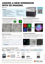
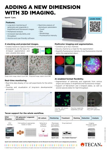
ADDING A NEW DIMENSION WITH 3D IMAGING. Spark® Cyto. LEARN MORE Features. • Long-term monitoring of spheroids and organoids in brightfield and fluorescent images • Multiplexed analysis • Increased reproducibility and throughput • Intuitive and user-friendly software • Real-time analysis of phenotypic parameters: -C ount -S ize - Morphology - Fluorescence intensity Z-stacking and projected images. Multicolor imaging and segmentation. • Create Z-stacks to capture information in all three dimensions • Z-projections are the basis for AI-based segmentation and save valuable disk space • Combine up to four channels • Use any channel as a mask for the segmentation • Quantify fluorescence intensity of any fluorescence channel within the segmented object Single stack image, object count 269 Projected image, object count 599 AI enabled format flexibility. Real-time monitoring. • Real-time data display of live-cell experiments for the entire plate • Tracking and visualization of long-term developmental processes • Segmentation of spheroids and organoids from various origins and shapes in brightfield and fluorescence images • Support of flat-bottom and U-shaped plates, as well as several special plates for high throughput Corning™ 96, Human colon, NucBlue™, CyQuant™ Dog liver organoids, brightfield, 24-well Tecan support for the whole workflow. iPSC banking 3D spheroid / organoid generation Cell seeding in appropriate media or matrix 3D culture Spheroid or organoid formation Monitoring Phenotype Drug treatment (growth, morphology, proliferation, marker expression) Spark Cyto Imaging End-point assays (live/dead, ATP) Spark Cyto multimode reader and Duo Digital Dispenser are for Research Use Only. Not for use in diagnostic procedures. © 2025 Tecan Trading AG, Switzerland, all rights reserved. For disclaimer and trademarks please visit www.tecan.com Spark Cyto
Open the catalog to page 1All Tecan catalogs and technical brochures
 TouchTools™
4 Pages
 HydroSpeed™
6 Pages
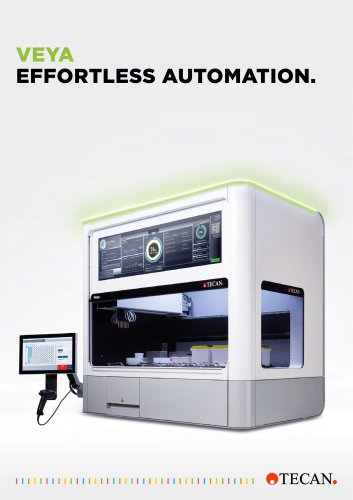 Veya®
Veya®8 Pages
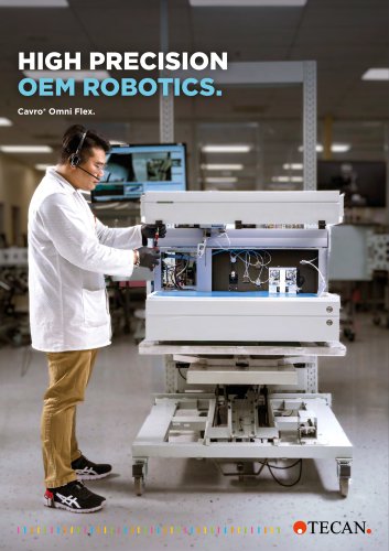 Cavro® Omni Flex
12 Pages
Archived catalogs
 Cavro ® Omni Flex
12 Pages
 Cavro ® robotics
12 Pages
 Te-Options
6 Pages
 FluentControl
6 Pages


- Solvent reagent
- Analysis software
- Dye reagent kit
- Sample preparation system
- Clinical chemistry reagent kit
- Automatic sample processor
- Microtiter plate
- Control software
- Quality control reagent
- Reporting software
- Laboratory software
- Windows software
- Laboratory sample preparation system
- Benchtop sample preparation system
- 96-well microplate
- Pipette tip
- Monitoring software
- Syringe pump
- Cloud-based software
- Automated software