
Catalog excerpts
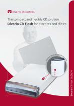
The compact and flexible CR solution Divario CR-Flash for practices and clinics
Open the catalog to page 1
3 models with different scanning speeds from
Open the catalog to page 2
Divario CR-Flash The compact CR system with scanning speed for every requirement With its robust cassettes, the Divario CR-Flash system provides a crystal-clear image quality for diagnostic imaging applications needed in small practices and hospitals. Thanks to its innovative design the reader can be placed on a counter or wall-mounted. Together with the professional image acquisition software dicomPACS®DX-R, the CR system offers all necessary image processing tools. The solution can be adapted to fit specific clinical purposes. It is also ideal as a secondary or backup system where there...
Open the catalog to page 3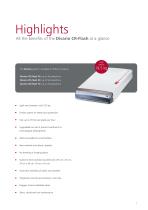
All the benefits of the Divario CR-Flash at a glance The Divario system is available in different versions: Divario CR-Flash 30: up to 30 plates/hour Divario CR-Flash 50: up to 50 plates/hour Divario CR-Flash 70: up to 70 plates/hour ■ Light and compact: only 19,5 kg ■ Fanless system for better dust protection ■ Fast: up to 70 full-size plates per hour ■ Upgradable on-site to protect investments in technological developments ■ Wall-mountable for small facilities ■ New resistant and robust cassettes ■ No bending of imaging plates ■ Suited to three standard cassette sizes (35 cm x 43 cm, ■...
Open the catalog to page 4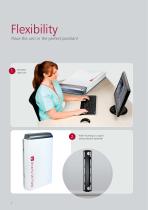
Flexibility Place the unit in the perfect position! Standard table unit Wall mounting as a spacesaving solution (optional)
Open the catalog to page 5
Software Benefits of the professional dicomPACS®DX-R X-ray acquisition software Modern graphical user interface (GUI) adaptable to almost any language Touchscreen operation - to ensure quick and efficient work and a smooth workflow Capture of patient data via DICOM Worklist, BDT/GDT, HL7 or other protocols - data may also be captured manually Use of DICOM Procedure Codes for the transfer of all relevant examination data directly from the connected patient management system (HIS/RIS) Freely configurable body parts with more than 200 projections and numerous possible adjustments in already...
Open the catalog to page 6
Benefits of flexible image acquisition The configurable generator interface enables the user to control X-ray generators or X-ray systems by different manufacturers, delivering the generator settings directly from the software Option for the parallel operation of a flat panel and a CR system included in the standard package. The user has the choice to take the next image with either the flat panel or the integrated CR system. This flexibility also provides an excellent emergency concept in case of a defect flat panel. Integration of dose area product meters (DAP) - the readings are saved...
Open the catalog to page 7
Operation of the acquisition software The correct settings for adults and children at a mouse click Job creation Marion the for t Char of an g nin plan dual i indiv y job a X-r Sw itc h to th e pl an ni ng of X - ra y jo bs fo r ch ild re n Radiographic positioning guide s an Show of a y ple exam t X-ra c corre age im Presentati on of helpful hints for the positioning of the patient, central beam, tips and tricks, frequent errors etc. 6
Open the catalog to page 8
Preview of the X-ray image and worklist of Preview rrent the cu image X-ray Generator control l a to r p a n e T h e ge n e r s all value d is p l a y s gs a n d s e tt in s As, focu (kVp, m ommended e tc . ) r e c e c if ic for a sp n e x a m in a ti o
Open the catalog to page 9
Image processing Automatic image processing for optimal quality Perfect images at all times - generally no adjustment required Integrated software for automatic image optimisation Professional, adaptable image processing for each individual examination to obtain best possible image settings for the needs of each customer Due to specially developed processes, the image processing allows the user to vary the X-ray settings on a large scale while the image quality remains virtually the same (possibility of reducing the dosage) Bones and soft tissue in one image - this enables the user to...
Open the catalog to page 10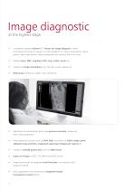
Image diagnostic at the highest stage Completely integrated dicomPACS® Viewer for image diagnosis, further processing and storage of images in an SQL database incl. image manipulations, export options, layout adjustments, freely configurable user interface and much more Stepless zoom, PAN, magnifyer, ROI, crop, rotate, mirror etc. Insertion of image annotations, e.g. free texts, arrows, ellipses etc. Measuring of distances, angles, areas and density Adjustment of window/level options and gamma correction, sharpening filters, noise suppression Many additional functions such as Chiro Tools,...
Open the catalog to page 11
Integrated viewer Completely integrated ® dicomPACS viewer for image diagnosis An integrated prosthesis documentation module provides preoperative planning (optional). The system enables fast and easy customisation of the operating interface for individual customer preferences.
Open the catalog to page 12
Useful tools such as the configurable measuring magnifier make diagnosis much easier. The stitching module merges a number of separate digital X-ray images into a single image. Comprehensive search tools enable the comparison of X-ray examinations of one or more patients.
Open the catalog to page 13
Mobile Web-based viewer dicomPACS®MobileView for mobile and stationary devices (optional) The web-based viewer dicomPACS®MobileView counts among the many extension modules of dicomPACS® diagnostic software. As a virtually independent browser, it allows the viewing of image material on mobile devices also outside a clinic or a practice. The doctor or the nursing staff can access all image material from the dicomPACS® system worldwide via a network connection. ® In addition to mere diagnostic evaluation of images, the dicomPACS MobileView viewer allows diagnostic reports to be captured and...
Open the catalog to page 14
Media IH-tDlilech Ml 11\Itwl/ Sfit) humw M Drama CH-F ' FtreCR 'iiiman The web-based viewer offers an important range of functions of a professional PACS viewer: ■ Draw lines and arrows (multi- different grids ■ Adjust brightness/ contrast ■ Flip and rotate images ■ Adjust brightness / contrast ■ Full screen, fit image ■ Scroll through image series ■ Cine loop for multi frame series ■ Export images and documents
Open the catalog to page 15All OR Technology - Oehm und Rehbein catalogs and technical brochures
-
Amadeo R motorised
5 Pages
-
Amadeo V-DR mini
6 Pages
-
XenOR 43CL
2 Pages
-
XenOR 35CW
2 Pages
-
4343F
2 Pages
-
Amadeo Z-DR
7 Pages
-
Toshiba detector FDX3543RPW
3 Pages
-
Beyond a good image
9 Pages
-
Digital X-ray imaging - Overview
11 Pages
-
GIERTH TR 90/20 Battery
2 Pages
-
JOB Porta 120 HF
1 Pages
-
JOB Porta 100 HF
1 Pages
-
Poskom PXM-40BTP
1 Pages
-
Poskom PXM-20BTP
1 Pages
-
Chiro Tools
4 Pages
-
Medici DR Systems vet
23 Pages
-
Medici DR Systems
28 Pages
-
GIERTH HF 400 ML
2 Pages
-
GIERTH TR 90/30
2 Pages
-
Veterinary X-Ray Systems
6 Pages
-
dicom PACS DX-R 19"
2 Pages
-
FLAATZ 560
2 Pages
-
FDX 4343R
2 Pages
-
dicom PACS®DX-R
2 Pages
-
DR flat panel upgrade kit
17 Pages
Archived catalogs
-
GIERTH TR 90/20 Battery1
2 Pages
-
Voxar 3DTM
12 Pages
-
MobileView
2 Pages


