
Catalog excerpts

X-ray Acquisition Software Acquisition and diagnostic software for X-ray images from DR flat panels or CR systems in human and veterinary medicine
Open the catalog to page 1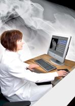
Image displa Multi monitor station
Open the catalog to page 2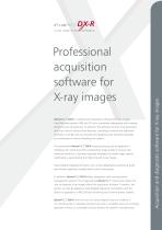
Professional acquisition software for X-ray images ® dicomPACS DX-R is a professional acquisition software for X-ray images from flat panel systems (DR) and CR units (computed radiography with imaging plates) by any manufacturer. In addition, the software controls X-ray generators and X-ray units of various manufacturers, providing a smooth and systematic workflow. A simple and user friendly GUI (graphical user interface) operated by touchscreen or mouse completes the system. ® The professional dicomPACS DX-R image processing can be adapted to individual user needs and offers outstanding...
Open the catalog to page 3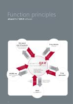
Flat panel and CR systems X-ray devices (motorised) dicomPACS klist Wor atient OMvery of pination DIC eli D ot Co o n co rise tro llim d l o at syst f th or em e et , c. (different manufacturers, also dental panel) X-ray Acquisition Software exam tions and instruc - operation software for generator and panel - image processing - image management n atio ave firm tions h Conn instruc out d e wh carrie n bee (Patient management system) Unless provided by HIS/RIS DICOM Worklist DICOM store Output of processed images incl. all patient and exposure data X-ray generator
Open the catalog to page 4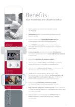
Benefits User friendliness and smooth workflow Modern graphical user interface (GUI) adaptable to almost any language Touchscreen operation – to ensure quick and efficient work and a smooth workflow Capture of patient data via DICOM Worklist, BDT/GDT, HL7 or other protocols – data may also be captured manually Remote control for X-ray units Use of DICOM Procedure Codes for the transfer of all relevant examination data directly from the connected patient management system (HIS/RIS) Freely configurable body parts with more than 400 projections and numerous possible adjustments in human and...
Open the catalog to page 5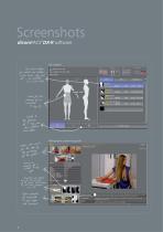
The correct settings for adults and children or for horses, dogs and cats – are available at a mouse click Steve Miller dicomPACS Chart for the planning of an individual X-ray job h so u n d V id e o w it p b y st e f o r th e ti o n in g st e p p o si e n t a ti o f th e p S h ow s an a ex am pl e of y co rr ec t X - ra im ag e Presentation of helpful hints for the positioning of the patient, central beam, tips and tricks, frequent errors etc. Opens examples of inaccurate X-ray images with comments X-ray Acquisition Software Radiographic positioning guide
Open the catalog to page 6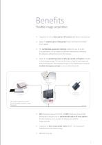
Benefits Flexible image acquisition Integration of various flat panel and CR systems by different manufacturers Option to connect up to 3 flat panels (bucky, wall stand and mobile) to one system The configurable generator interface enables the user to control X-ray generators or X-ray systems by different manufacturers, delivering the generator settings directly from the software Option for the parallel operation of a flat panel and a CR system included in the standard package. The user has the choice to take the next image with either the flat panel or the integrated CR system. This...
Open the catalog to page 7
The professional dicomPACS DX-R image processing Perfect images at all times – generally no adjustment required Integrated software for automatic image optimisation Professional, adaptable image processing for each individual examination to obtain best possible image settings for the needs of each customer Due to specially developed processes, the image processing allows the user to vary the X-ray settings on a large scale while the image quality remains virtually the same (possibility of reducing the dosage) Bones and soft tissue in one image – this enables the user to significantly...
Open the catalog to page 8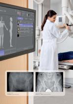
Exposure with standard image processing Exposure with dicomPACS®DX-R image processing
Open the catalog to page 9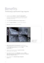
Benefits Outstandingly sophisticated image diagnosis Completely integrated dicomPACS® viewer for image diagnosis, further processing and storage of images in an SQL database incl. image manipulations, export options, layout adjustments, freely configurable user interface and much more Stepless zoom, PAN, magnifyer, ROI, crop, rotate, mirror etc. Insertion of image annotations, e.g. free texts, arrows, ellipses etc. Measuring of distances, angles, areas and density dicomPACS viewer for image diagnosis Special purpose tools for the veterinarian (Specialised filters for the optimised depiction...
Open the catalog to page 10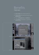
Benefits Image export Export of images to JPEG, TIFF, BMP and DICOM formats Printing of images both on Windows printers and laser imagers via DICOM Basic Print Creation of DICOM patient CDs with free WEB viewer Inbuilt e-mail tool to image distribution - no external Email application necessary E-mail tool Image print
Open the catalog to page 11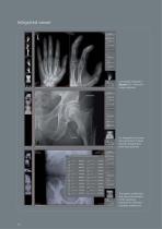
Integrated viewer Completely integrated ® dicomPACS viewer for image diagnosis An integrated prosthesis documentation module provides preoperative planning (optional). The system enables fast and easy customisation of the operating interface for individual customer preferences.
Open the catalog to page 12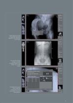
Useful tools such as the configurable measuring magnifier make diagnosis much easier. The stitching module merges a number of separate digital X-ray images into a single image. Comprehensive search tools enable the comparison of X-ray examinations of one or more patients.
Open the catalog to page 13
Cloud-based Digital access and archiving of images and diagnostic reports via Internet ORCA - the Cloud-based archive and teleradiology solution by OR Technology Even for state-of-the-art practices and hospitals, the rapidly rising data flood of digital images, diagnostic reports and other documents is becoming increasingly challenging. Current legislation demands safe and long-term storage of patient data which generally requires investing in expensive hardware infrastructure as well as maintenance and corresponding staff costs. To this end, we developed the ORCA Cloud archiving solution,...
Open the catalog to page 14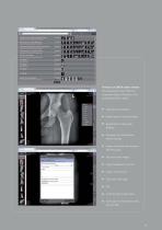
Features of 0/7CA online viewer: The web-based viewer offers an important range of functions of a professional PACS viewer: Drawing Lines and Arrows Image comparison by choosing different grids Flip and rotate images Adjust brightness/ contrast Full screen, fit image Scroll through image series Cine loop for multi-frame series
Open the catalog to page 15All OR Technology - Oehm und Rehbein catalogs and technical brochures
-
Amadeo R motorised
5 Pages
-
Amadeo V-DR mini
6 Pages
-
XenOR 43CL
2 Pages
-
XenOR 35CW
2 Pages
-
4343F
2 Pages
-
Amadeo Z-DR
7 Pages
-
Toshiba detector FDX3543RPW
3 Pages
-
Beyond a good image
9 Pages
-
Digital X-ray imaging - Overview
11 Pages
-
GIERTH TR 90/20 Battery
2 Pages
-
JOB Porta 120 HF
1 Pages
-
JOB Porta 100 HF
1 Pages
-
Poskom PXM-40BTP
1 Pages
-
Poskom PXM-20BTP
1 Pages
-
Chiro Tools
4 Pages
-
Medici DR Systems vet
23 Pages
-
Medici DR Systems
28 Pages
-
GIERTH HF 400 ML
2 Pages
-
GIERTH TR 90/30
2 Pages
-
Veterinary X-Ray Systems
6 Pages
-
dicom PACS DX-R 19"
2 Pages
-
FLAATZ 560
2 Pages
-
FDX 4343R
2 Pages
-
dicom PACS®DX-R
2 Pages
-
DR flat panel upgrade kit
17 Pages
Archived catalogs
-
GIERTH TR 90/20 Battery1
2 Pages
-
Voxar 3DTM
12 Pages
-
MobileView
2 Pages


