
Catalog excerpts

Digital image processing Digital image processing with dicomPACS®vet
Open the catalog to page 1
dicomPACS®vet will make your dream of a paperless veterinary practice come true. All images as well as any type of document (e.g. diagnostic reports, records of healing processes, faxes) are stored by dicomPACS vet ® in a digital patient file and can be accessed immediately with a simple mouse click. Well designed archiving and backup solutions guarantee fast access to all data while observing the highest security standards in accordance with the internationally recognised guidelines for human medicine. In addition, dicomPACS vet can be integrated easily with all the popular practice ®...
Open the catalog to page 3
Benefits of dicomPACS®vet at one glance Full diagnostic software for all workstations in your practice (no 'light' versions) User friendly and clearly arranged structure, minimal training requirements and short familiarisation period Individual adjustment of the user interface to your field of specialisation and individual requirements Flexible allocation of shortcut keys for many functions to allow fast work without a mouse Parallel processing (e.g. option to continue working during a CD burning process) Permanent online availability of all images and data in the network – no need to store...
Open the catalog to page 4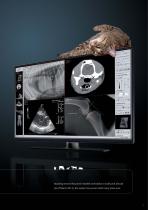
Boasting several thousand installed workstations locally and abroad (as of March 2013), the system has proven itself many times over.
Open the catalog to page 5
Services offered Integrated modules and tools Perfect integration of all imaging devices into your existing computer network is an important condition for a smooth and reliable workflow. Apart from X-ray systems, a wide range of devices including ultrasound, endoscopy, fluoroscopy, CT and MRI systems as well as digital cameras can be connected. In addition to imaging devices, you can also store documents such as faxes and letters digitally in the digital patient file of your practice management system. With dicomPACS®vet, all data is immediately available and can even be easily forwarded on...
Open the catalog to page 6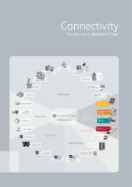
Connectivity The diversity of dicomPACS vet ® X-ray scanner Dental vet X-ray CR/ DR Operation documentation Image sources X-ray units Document scanner Leonardo DR suitcase solution Video projector Image output Laser printer Laser imager Viewing station Amadeo complete DR system Solutions of OR Technology incl. acquisition and diagnostic software Image archiving Image viewing Divario CR solution with cassettes Image processing Multimonitor workstation Archive server Interfaces to practice management system Home workstation Diagnostic station Telemedicine/ Cloud
Open the catalog to page 7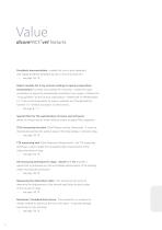
dicomPACS®vet features Prosthesis documentation - enables the user to plan operations with digital prosthesis templates by one or more manufacturers see page 18/ 19 Report module for X-ray services relating to equine prepurchase examinations [currently only available for Germany] - enables the quick compilation of reports by automatically assembling X-ray images. It follows the “X-ray guideline” by the German organisations “Gesellschaft für Pferdemedizin e.V.” (non-profit organisation for equine medicine) and “Bundestierärztekammer e.V.” (Federal association of veterinarians). see page 8 -...
Open the catalog to page 8
Statistics Module - enables freely configurable analysis of the complete database Video Modules - enable standard and non-standard video signals to be recorded as single images and video sequences Web Server - enables image distribution within the hospital or to referring doctors via the internet and guarantees very fast image accessibility in original quality (DICOM) see page 14 Processing of CT and MRI series - dicomPACS®vet includes professional tools such as MPR and MIP to evaluate cross section series see page 16/ 17 Hanging protocols Telemedicine Special solution for multiple archives...
Open the catalog to page 9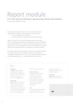
Report module for X-ray services relating to equine prepurchase examinations [currently only available for Germany] Presale and prepurchase examinations for horses are always particularly challenging for veterinarians. Such specialised examinations must be carried out swiftly yet very meticulously documented very well, in great detail and extremely consistently. After all, the owner of the animal justifiably expects optimal service when it comes to undertaking the examination and presenting the results in a professional and comprehensible fashion. Since administrative work is bothersome yet...
Open the catalog to page 10
Joe eioqqs, Dcmc hgrser -01.04.1999 Equine Clinic Veterinarian specializing in horses X-ray protocol for prepurchase examinations UELN number: Prepurchase examination number: PE-4163786 Left front 1.4.1: measured at right angle/ middle of Pedal bone < 1,5 cm - with different interpretation Left front 2.2.4: contour margo solearis - irregular ; 2.3.3: aligned - difference left from right X-ray protocol
Open the catalog to page 11
Workflow for a prepurchase horse examination 2. Start report module Current image assign 4. Display diagnostic report options left front Foot right front Foot Current image assign left hind left front Foot right front Foot left hind Foot Knee Knee left right left right Knee Knee Projection add or delete Assignment cancel back Back Projection add or delete Assignment cancel 5. Diagnostic report/ evaluation Create report
Open the catalog to page 12
By means of a single click, the completed prepurchase examination ® report is stored together with the examination images in dicomPACS vet, where it can be accessed again in the original version at any point in time.
Open the catalog to page 13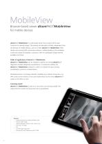
Browser-based viewer dicomPACS®MobileView for mobile devices dicomPACS®MobileView is a web-based viewer, that contains all the basic functions for viewing images. The viewing can take place virtually independent from the browser on mobile devices, such as an iPad. dicomPACS MobileView offers ® doctors and nursing staff a previously unknown, mobile freedom in the workplace inside and outside of hospitals or practices, with the radiological image material available at all times. Fields of application of dicomPACS MobileView ® dicomPACS MobileView can be installed in addition to existing...
Open the catalog to page 14All OR Technology - Oehm und Rehbein catalogs and technical brochures
-
Amadeo R motorised
5 Pages
-
Amadeo V-DR mini
6 Pages
-
XenOR 43CL
2 Pages
-
XenOR 35CW
2 Pages
-
4343F
2 Pages
-
Amadeo Z-DR
7 Pages
-
Toshiba detector FDX3543RPW
3 Pages
-
Beyond a good image
9 Pages
-
Digital X-ray imaging - Overview
11 Pages
-
GIERTH TR 90/20 Battery
2 Pages
-
JOB Porta 120 HF
1 Pages
-
JOB Porta 100 HF
1 Pages
-
Poskom PXM-40BTP
1 Pages
-
Poskom PXM-20BTP
1 Pages
-
Chiro Tools
4 Pages
-
Medici DR Systems vet
23 Pages
-
Medici DR Systems
28 Pages
-
GIERTH HF 400 ML
2 Pages
-
GIERTH TR 90/30
2 Pages
-
Veterinary X-Ray Systems
6 Pages
-
dicom PACS DX-R 19"
2 Pages
-
FLAATZ 560
2 Pages
-
FDX 4343R
2 Pages
-
dicom PACS®DX-R
2 Pages
-
DR flat panel upgrade kit
17 Pages
Archived catalogs
-
GIERTH TR 90/20 Battery1
2 Pages
-
Voxar 3DTM
12 Pages
-
MobileView
2 Pages


