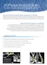
AQPlaque Analysis Suite

AQPlaque Analysis Suite
AQPlaque provides advanced tools for the visualization and evaluation of atherosclerotic plaque. It allows for the characterization of plaque constitution and morphology, including wall remodeling, luminal encroachment, and density/intensity distribution of CT/MR values within the plaque.
Clinical Evaluations
A case study by C. Herzog et al. at Johann Wolfgang Goethe-University compared MDCT, MR, and histopathology for detecting and characterizing atherosclerotic lesions using AQPlaque. The study found MDCT superior for lesion detection, while MR was better for sub-characterization. Observers reported excellent agreement using the software tools.
Integration and Capabilities
AQPlaque integrates with luminal analysis tools for stenosis calculations, tissue type identification, and vessel delineation. It calculates diameters, areas, and stenosis percentages, and exports images to DICOM, CD, or local PCs. The tools are part of the optional Vascular analysis package of the Aquarius Workstation.
Visualization and Measurement Tools
For CT images, HU intensity ranges can be mapped to color overlays for lesion visualization. Flexible region-of-interest tools exclude non-vascular structures, and the volume of each tissue category is automatically calculated. Stenosis measurement can use three-point QCA or simple-ratio logic.
Advanced Analysis Features
The Aquarius Workstation provides flexible tools for defining lumen and wall boundaries. The AQPlaque histogram tool plots HU or intensity values against voxel count within the ROI. Quartile volume calculation within the ROI is automatic, and lumen or whole-vessel area can be compared with intravascular ultrasound (IVUS).
Catalog excerpts
AQPlaque Analysis Suite Enhanced Plaque Visualization and Evaluation Improved Vascular & Cardiac Plaque Visualization & Evaluation AQPlaque offers a suite of tools for the evaluation and visualization of atherosclerotic plaque. Parameters relating to plaque constitution and morphology, including the overall extent of wall remodeling, luminal encroachment, and the density/intensity distribution of CT/MR values within the plaque, can be characterized with the AQPlaque tools. Reported Clinical Evaluations of AQPlaque In a case study performed by C. Herzog et. al. at the Johann Wolfgang Goethe-University, Frankfurt (Clinical Case Studies in the Third Dimension, Volume 2, 2006, TeraRecon, Inc.), investigators reported a comparison between MDCT, MR, and histopathology for the detection and characterization of atherosclerotic lesions, using the AQPlaque suite. The results suggested that MDCT is superior to MR for detection of such lesions, while MR is superior to MDCT for further sub-characterization of lesion type. The case study reported excellent agreement between observers applying the software tools. AQPlaque Features • AQPlaque tools are integrated with luminal analysis tools for stenosis calculations. • Identify and quantify various tissue types from CT and MR images. • Delineate vessels' outer and inner walls from surrounding tissue and lumen. • Calculate diameters, areas, and percentage of stenosis. • Export images to DICOM, CD, or to a local PC.
Open the catalog to page 1
AQPlaque Analysis Suite Enhanced Plaque Visualization and Evaluation Aquarius w o r k s t a t i o n • AQPlaque tools are integrated into the optional Vascular Analysis package of the Aquarius Workstation. • For CT images, a set of HU intensity ranges can be defined and mapped to a color overlay to visualize various elements of a lesion. • Flexible region-of-interest tools are available to exclude non-vascular structures. • The volume of each defined tissue category is automatically calculated within the region of interest. • Stenosis measurement may be based on three-point QCA or simple-ratio...
Open the catalog to page 2All TeraRecon catalogs and technical brochures
 iNtuition Brochure
20 Pages
 iNteract+
4 Pages
 PACS Partnership Brochure
8 Pages
 ? iNtuition SHARE Brochure
6 Pages
 iNtuitionEMV Brochure
6 Pages
 iNtuition TAVI Brochure
6 Pages

