Use of TissuePatchDural? for Dural closure for revision craniotomy
Use of TissuePatchDural? for Dural closure for revision craniotomy
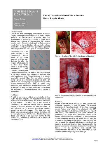
A 33-year-old female presented with a large right posterior frontal low-grade glioma in 2010. She underwent an awake craniotomy and tumor resection in March 2011, using TissuePatchDural™ for dural closure. Histology confirmed a Diffuse Astrocytoma, WHO Grade II. Follow-up showed a potential tumor recurrence, leading to plans for revision surgery.
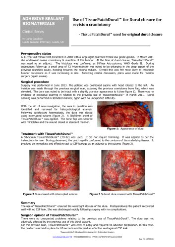
The revision surgery took place in June 2013. The patient was positioned supine, and the previous surgical scar was incised to expose the craniotomy bone flap. The dura appeared intact with a granular texture. Neuronavigation aided in identifying and removing the area for histopathological analysis. The dura was closed with interrupted sutures, and a 50x50mm TissuePatchDural™ was applied. The bone flap was secured with miniplates, and the wound was closed.
A 50×50mm TissuePatchDural™ was used without trimming, conforming rapidly to tissue contours and providing an effective seal against CSF leakage.
TissuePatchDural™ ensured watertight dural closure. The patient recovered well postoperatively with no CSF leak and was discharged without complications.
No issues arose from previous TissuePatchDural™ use. The product was easy to apply, requiring no preparation, and effectively sealed against CSF leak.
Catalog excerpts

ADHESIVE SEALANT BIOMATERIALS Use of TissuePatchDural™ for Dural closure for revision craniotomy Clinical Series -‐ TissuePatchDural™ used for original dural closure Mr John Goodden Leeds General Infirmary, Leeds, UK Pre-operative status A 33-year-old female first presented in 2010 with a large right posterior frontal low grade glioma. In March 2011 she underwent awake craniotomy & resection of this tumour. At the time of dural closure, TissuePatchDural™ was used as an adjunct. The histology was confirmed as Diffuse Astrocytoma, WHO Grade II. During subsequent follow-up, a small area of T2 hyperintensity was noted to be enlarging in the deep aspect of the previous resection cavity, heading towards the corona radiata. Overall this was felt most likely to represent tumour recurrence as it was increasing in size. Following careful discussion, plans were made for revision surgery (again awake). Surgical procedure Surgery was performed in June 2013. The patient was positioned supine with head rotated to the left. An incision was made through the previous surgical scar, exposing the previous craniotomy bone flap, which was elevated. The dura was noted to be intact with a slightly granular appearance to it (see figure 1). There was no evidence of excessive scarring in relation to the previous use of TissuePatchDural™ in March 2011. Dural opening was performed in standard manner, again with no unexpected difficulty. With the aid of neuronavigation, the area in question was identified and removed for histopathological analysis. Following satisfactory haemostasis, the dura was closed using interrupted sutures (figure 2). A 50x50mm sheet of TissuePatchDural™ was applied. The bone flap was secured with miniplates and the wound closed in standard manner. Figure 1: Appearance of dura Treatment with TissuePatchDural™ A 50×50mm TissuePatchDural™ (TD-02) was used. It did not require trimming. It was applied as per the instructions for use. During placement, the patch rapidly conformed to the contours of the underlying tissues. It provided an immediate and effective seal to CSF leakage as an adjunct to the sutures (figure 3). Figure 2 Dura closed with interrupted sutures Figure 3 Sutured dura covered with TissuePatchDural™ Summary The use of TissuePatchDural™ ensured the watertight closure of the dura. Postoperatively the patient recovered well with no CSF leak. She was discharged rapidly following surgery with no complications. Surgeon opinion of TissuePatchDural™ There were no unexpected problems relating to the previous use of TissuePatchDural™. The dura was not adversely affected by the previous use of this dural sealant. For this revision case, TissuePatchDural™ was easy to apply and required no advance preparation. In this case, the product was held in place for 60 seconds and formed an effective seal against CSF leak. Tissuemed Ltd.•5 Killingbeck Drive•Leeds•LS14 6UK•United Kingdom www.tissuemed.com•Tel +44(0)1132000500•Fax +44(0)1132407343•©Tissuemed 2013 July 2013 CS69/01
Open the catalog to page 1All Tissuemed catalogs and technical brochures
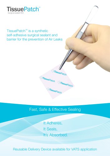
 TissuePatch
2 Pages