Use of TissuePatchDural? in a Porcine Dural Repair Model
Use of TissuePatchDural? in a Porcine Dural Repair Model
The document discusses the challenges of cerebrospinal fluid (CSF) leakage in cranial surgery and introduces TissuePatchDural, a CE-marked product designed to effectively seal the Dura Mater. It highlights the product's advantages such as low material bulk, rapid delivery, zero preparation time, and short application time. TissuePatchDural is a sterile, absorbable film that is absorbed in about 50 days.
Methods
The study involved six porcine subjects. A craniotomy was performed on each side of the midline, and a dural defect was created. The control side was sutured, while the test side was sutured and treated with TissuePatchDural.
Results
In the control group, two sites required additional intervention due to CSF leaks, while TissuePatchDural was 100% effective in preventing CSF leakage. At 14 and 28 days, all animals showed no neurological changes, and all dural defects were sealed. The TissuePatchDural sites showed minimal inflammatory response compared to the control.
Conclusion
The study concludes that TissuePatchDural is safe and effective in reducing CSF leaks during neurosurgical procedures. It performed well intraoperatively, sealing all leaks, and showed predictable degradation and foreign body response.
Catalog excerpts

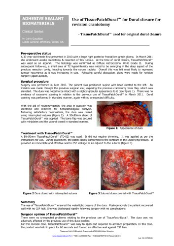
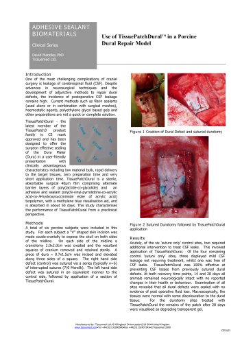
ADHESIVE SEALANT BIOMATERIALS Clinical Series Use of TissuePatchDuralTM in a Porcine Dural Repair Model David Mandley PhD Tissuemed Ltd. One of the most challenging complications of cranial surgery is leakage of cerebrospinal fluid (CSF). Despite advances in neurosurgical techniques and the development of adjunctive methods to repair dural defects, the incidence of postoperative CSF leakage remains high. Current methods such as fibrin sealants (used alone or in combination with surgical meshes), haemostatic agents, polyethylene glycol based gels and other preparations are not a quick or complete solution. TissuePatchDural - the latest member of the TissuePatch3 product family is CE mark approved and has been designed to offer the surgeon effective sealing of the Dura Mater (Dura) in a user-friendly presentation with clinically advantageous characteristics including low material bulk, rapid delivery to the target tissues, zero preparation time and very short application time. TissuePatchDural is a sterile, absorbable surgical 40µm film comprising alternate barrier layers of poly(lactide-co-glycolide) and an adhesive and sealant poly(N-vinyl-pyrrolidone-co-acrylic acid-co-N-hydroxysuccinimide ester of acrylic acid) terpolymer, with a methylene blue visualisation aid, and is absorbed in about 50 days. This study characterises the performance of TissuePatchDural from a preclinical perspective. A total of six porcine subjects were included in this study. For each subject a “V” shaped skin incision was made caudo-cranially to expose the skull on both sides of the midline. On each side of the midline a craniotomy 2.0x2.0cm was created and the resultant squares of cranium removed and retained sterile. A piece of dura ≈ 0.7x1.5cm was incised and elevated along three sides of a square. The right hand side defect (control) was sutured via a series (typically n=6) of interrupted sutures (7/0 Mersilk). The left hand side defect was sutured in an equivalent manner to the control side, followed by application of a section of TissuePatchDural. Figure 1 Creation of Dural Defect and sutured durotomy Figure 2 Sutured Durotomy followed by TissuePatchDural application Acutely, of the six ‘suture only’ control sites, two required additional intervention to treat CSF leaks. This involved application of TissuePatchDural. Of the four remaining control ‘suture only’ sites, three displayed mild CSF leakage not requiring treatment, whilst one was free of CSF leaks. TissuePatchDural was 100% effective at preventing CSF losses from previously sutured dural defects. At both recovery time points, 14 and 28 days all animals remained neurologically intact with no reported changes in their health or behaviour. Examination of all sites revealed that all dural defects were sealed with no evidence of post operative fluid loss. Macroscopically, the tissues were normal with some discolouration to the dural tissue. For the durotomy sites treated with TissuePatchDural the remains of the patch after 28 days were visualised as degrading transparent gel.
Open the catalog to page 1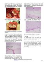
Qualitatively (by appearance and manipulation) the ‘suture only’ sites were reported as slightly more fibrosed (thicker) when compared to the ‘suture + TissuePatchDural’ treated durotomies. There were no other remarkable features evident during the course of the macroscopic examination. exception of one durotomy, in which there was pre-existing damage to the brain, the brain beneath the TissuePatchDuraltreated Dura displayed minimal changes and there was no evidence of damage to the underlying brain adjacent to or remote from the dural repair. Figure 3 TissuePatchDural Durotomy site – 28 days...
Open the catalog to page 2All Tissuemed catalogs and technical brochures
 TissuePatch
2 Pages