
Catalog excerpts

Maestro2 Optical Coherence Tomography True Color Fundus Camera ONE SCAN. ONE REPORT. ONE INSTRUMENT. The fast, one-touch, automated OCT and Fundus Camera. * OCTA optional extra in some countries. Please check with the distributor in your country.
Open the catalog to page 1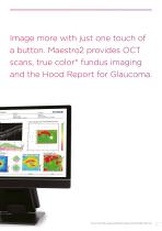
Image more with just one touch of a button. Maestro2 provides OCT scans, true color* fundus imaging and the Hood Report for Glaucoma. *True, full color fundus image simultaneously captured with white light, 24-bit color.
Open the catalog to page 3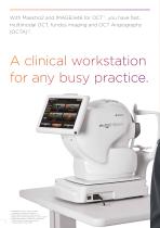
With Maestro2 and IMAGEnet6 for OCT*1, you have fast, multimodal OCT, fundus imaging and OCT Angiography (OCTA)*2. A clinical workstation for any busy practice. *1 MAGEnet6 for OCT is the standard I component software for Maestro2. *2 CTA optional extra in some countries. O Please check with the distributor in your country. *3 pplicable distance is subject to the device’s A communication performance and the communication environment.
Open the catalog to page 4
Maestro2 offers RTC (Remote Tablet Control) for social distance protocol.*3
Open the catalog to page 5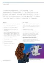
Introducing automated OCT, true color* fundus photography and automated OCT Angiography in one compact instrument. With the touch of a button, OCTA provides you instantaneous vascular structure information - from our world-renowned, multimodal OCT solution. Features: • OCT and true color* fundus photography A user-friendly OCT. The Maestro2 uses robotic technology and improves practice efficiency whilst providing optimal patient care. • Fully automated image capture • Compact and space saving design • wide scan with Hood Report for 3D Glaucoma • Reference database comparison for full...
Open the catalog to page 6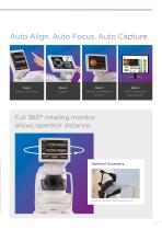
Auto Align. Auto Focus. Auto Capture. Step 1 Select a scan type. Step 3 Results are displayed instantly. Step 4 Report displayed automatically. Full 360° rotating monitor allows operator distance. Optional Accessory Anterior segment attachment (HA-2)
Open the catalog to page 7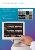
Introducing fully automated OCT Angiography*1 At the touch of a button, Maestro2 provides instantaneous vascular flow information without the need for contrast dye injection, together with comprehensive segmentation to enable advanced diagnosis. OCT Angiography includes OCTA Density.*2 *1 OCTA optional extra in some countries. Please check with the distributor in your country. *2 The OCTA Density is defined as the ratio between the high signal area and low signal area and it is displayed in color and/or number.
Open the catalog to page 8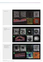
Diabetic Retinopathy (DR) PinPoint™ Registration of microaneurysms*3 *1 Michael H. Chen, OD *2 Prof. Siamak Ansari Shahrezaei, MD PhD (Karl Landsteiner Institute for Retinal Research and Imaging) *3 Miho Nozaki, MD, PhD (Nagoya City University Hospital)
Open the catalog to page 9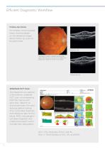
Efficient Diagnostic Workflow Follow-Up Scans For smaller, more localized areas, tracking based on the reference image allows follow-up scans to be performed. Baseline visit Tracking is used to capture exactly the same area at each visit and is available for single line, radial or 5 line cross scans. Follow-up visit Widefield OCT Scan The Maestro2 can capture a 12mmx9mm widefield OCT scan, encompassing both the macula and optic disc. Ideal for an annual eye exam, the scan reduces patient testing time. It provides thickness and reference data for the retina, RNFL and ganglion cell layers...
Open the catalog to page 10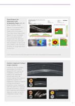
Hood Report for Glaucoma with Probability Maps with 3D Wide 12x9mm Scan Retinal Thickness/RNFL/ GCL and probability maps, all in one report. The New Hood Glaucoma Report is now available. This innovative report streamlines the decisionmaking process through the correlation of structure (GCL/RNFL) with function (overlay of visual field test locations).* *Donald C. Hood PhD, Translational Vision Science & Technology No.6 Vol.3 2014: Evaluation of a One-Page Report to Aid in Detecting Glaucomatous Damage. Anterior Segment Caliper/ Angle Analysis* Maestro2 has the added advantage of Anterior...
Open the catalog to page 11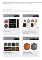
Extensive Set of Reports: Guidance for Diagnosis Extensive Set Of Reports Maestro2 provides rich analysis functions for the macular and disc regions and optic nerve. Comprehensive, predefined reports can be auto exported, quickly printed or sent to your image management system or EHR in common file formats. 5 Line Cross Report 5 line cross scan (6mm, 9mm) both horizontal and vertical in an instant. OCT Angiography Report Various OCTA scan protocols are available; 3x3mm, 4.5x4.5mm and 6x6mm. 3D Macula OU Report 3D Macula report available for single or both eyes if OU comparison is preferred....
Open the catalog to page 12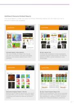
Additional Glaucoma Related Reports The Hood Report is often the report of choice following capture of a 3D wide scan but a choice of reports are available. 3D Wide Report (12mmx9mm) This scan provides an image of the macula and optic nerve head in one report, providing thickness and reference data for GCL+, GCL++ and RNFL. Glaucoma Analysis Report - Macula Based on the 3D Macula Vertical scan, this report provides RNFL, GCL+ and GCL++ thickness maps, comparison with reference data and symmetry analysis. 3D Disc Report OU Combines disc topography, fundus photography and RNFL thickness...
Open the catalog to page 13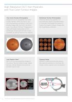
High Resolution OCT, Non-Mydriatic, and True Color Fundus Images True Color Fundus Photography* Maestro2 has an integrated true color fundus camera. With one touch, you can simultaneously acquire a true color fundus image with your OCT or OCTA scan. This allows PinPoint™ Registration and multimodal observation of the pathology. Small pupil function is also available, as well as fundus only capture. *Image courtesy: Michael H. Chen, O.D. Live Fundus View™ OCT-LFV is a live projection image of the retina. The live fundus image makes the disc, retinal vessels and scanning position easy to see....
Open the catalog to page 14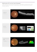
High Resolution OCT and Color Fundus Photography A high-resolution B-scan facilitates the evaluation of pathology by visualizing layers of the retina in fine detail. OCT B-scans, complemented by true color fundus photos, are vital for confident diagnosis. Healthy Eye* *Image courtesy: Michael H. Chen, O.D. Glaucoma* *Image courtesy: Michael H. Chen, O.D. Diabetic Retinopathy (DR)* *Image courtesy: Miho Nozaki, MD, PhD (Nagoya City University Hospital)
Open the catalog to page 15All Topcon Healthcare catalogs and technical brochures
-
 HARMONY
7 Pages
-
 OMS-800 Series
8 Pages
-
 Aladdin
11 Pages
-
 CV-5000PRO
10 Pages
-
 SOLOS
SOLOS4 Pages
-
 CL-300
6 Pages
-
 TRK-2P
8 Pages
-
 KR-1W
KR-1W7 Pages
-
 KR-1
KR-18 Pages
-
 KR-800S
10 Pages
-
 KR-800PA
2 Pages
-
 KR-800A
6 Pages
-
 KR-800/RM-800
4 Pages
-
 Chronos
8 Pages
-
 ACP-8
ACP-84 Pages
-
 CC series
5 Pages
-
 ATE Series
10 Pages
-
 FS-1 series
4 Pages
-
 IS-1 Series
16 Pages
-
 IS-600 III
12 Pages
-
 IS-100
8 Pages
-
 SP-1P
SP-1P8 Pages
-
 Henson 9000
9 Pages
-
 CT-1P
CT-1P10 Pages
-
 CT-800A
6 Pages
-
 CT-800
4 Pages
-
 MYAH
MYAH8 Pages
-
 CA-800
9 Pages
-
 Topcon Slit Lamp Series
7 Pages
-
 TRC-50DX
5 Pages
-
 Signal
2 Pages
-
 TRC-NW8 series
8 Pages
-
 TRC-NW400
8 Pages
-
 DRI OCT Triton™ Series
24 Pages
Archived catalogs
-
 VT-10 Vision Tester
4 Pages
-
 VT-10
VT-104 Pages