

2020 NEW

2020 NEW
The document provides detailed technical specifications and features of various dental imaging products, including CMOS intraoral sensors, phosphor plate scanners, and X-ray generators. These products are designed to capture high-quality digital images for dental diagnostics and treatment planning.
CMOS Intraoral Sensor - I-View Gold
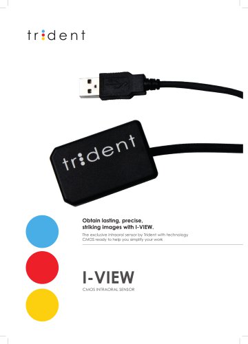
The I-View Gold sensor offers advanced technology for capturing intraoral images with consistent quality under various conditions. It features a CMOS detector, pixel size of 18 μm, and a theoretical resolution of 27 lp/mm. The sensor is available in two sizes with dimensions of 25.4 x 36.8 mm and 31.3 x 42.9 mm, and a thickness of 4.8 mm. It supports direct USB connection and is compatible with multiple platforms.
Deep View 4® Software
This software manages multiple databases and facilitates easy configuration for image acquisition. It includes advanced image filters, customizable templates for full mouth series exams, and options for voice guidance and DICOM format import/export.
Phosphor Plate Scanner
The scanner supports scanning, erasing, and reusing intraoral plates from size 0 to size 3. It offers high-definition images with a simple process and features like automatic voltage transformer and exposure time settings.
Intraoral X-Ray Generator
The RiX-70 DC and AC models provide high-frequency X-ray generation with features like low dose settings, high heat capacity, and extendable arms. The RiX-70 DC model operates at 60, 65, or 70 kV with a 0.4 mm focal spot.
X-View Imaging System
X-View offers a range of digital imaging devices for 2D and 3D imaging, supporting diagnostics and treatment planning. It includes features like pulsed radiation, high spatial resolution, and compatibility with DICOM 3.0.
Software and Compatibility
The Deep-View software suite provides dynamic and user-friendly imaging management with TWAIN options and DICOM compatibility. It supports multiuser environments and integrates with other dental management software through a bridge function.
Applications
The products are applicable in various dental fields, including implantology, endodontics, periodontology, orthodontics, and maxillofacial surgery. They assist in diagnosing TMJ disorders, bone morphology evaluation, infectious pathologies, and more.
Catalog excerpts

I-View Gold Technical information CMOS INTRAORAL SENSOR INTRAORAL X-RAY GENERATOR The most advanced technology to obtain intra-oral images. The sensor covers a wide range of exposure setting, capturing a consistent and diagnosable image under any conditions. TWAIN Interface DICOM files format Supports up to 10 network users “Bridge” function included Tube Voltage Anodic current Duty cycle Nominal power Total filtration Focal spot Distance focus to skin X Ray beam diameter at 200 mm focus Beam limiting device (additional) Reach Dimensions Size 1: 25.4 x 36.8mm Size 2: 31.3 x 42.9mm Thickness: 4.8 mm Size 1 20 x 30 mm active area Specifications Detector: CMOS Pixel size: 18 μm Theoretical Resolution: 27 lp/mm Dynamic Range: 12 bit Deep View 4® Software Latest generation of Deep View software with management of multiple databases and easy configuration of the device for image acquisition. Multiplatform option Size 2 26 x 36 mm active area Import / export of images in DICOM formats Print images with design customization Operative Mode Tube Voltage Anode Current Focal Spot I-View Sensitive (active) area Dimensions Number of Pixels Pixel size Spatial resolution Dynamic range Scintillator type I-View Gold 30 x 20 mm (size 1) 33 x 25 mm (size 2) 25 x 39 x h 5.3 (size 1) 30.5 x 42 x h 5.6 (size 2) 1500 x 1000 (size 1) 1650 x 1250 (size 2) 20 x 20 µm 20 lp/mm typical (theoretical 25 lp/mm) 14-bit ADC CsI 30 x 20 mm (size 1) 38 x 26 mm (size 2) 25.4 x 36.8 x 4.8 mm (size 1) 31.3 x 42.9 x 4.8 mm (size 2) 1666 x 1110 (size 1) 1998 x 1442 (size 2) 18 x 18 µm 22 lp/mm typical (theoretical 27 lp/mm) 12-bit ADC CsI INTRAORAL SENSOR PHOSPHOR PLATE SCANNER Sensor Pixel size Pixel/Voxel size 166 micron CBCT 100 micron PAN Acquisition Exposure time Scan time Reader Input Voltage Power Consumption Dynamic Range Resolution Connection to PC Plate size Measures Weight 100-240V AC, 50-60 Hz 24 VA 16 bit 14 lp/mm USB 2 (A type) cable 1 mt 0–1–2–3 170 x 190 x h300 mm 7 kg Optional Exams for 2DPAN and 3DPAN PC included Volumetric 3D Dentition Standard Panoramic 3D and 2D Sinuses Panoramic for Children Volumetric 3D Dentition 3D and 2D TMJ Frontal Dentition Left/Right TMJ Sections TMJ Sections 3D Maxillary Sinuses Standard Panoramic Maxillary Sinuses Panoramic for Children Frontal Dentition LL CEPH 30 x 24 LL CEPH 24 x 24 AP CEPH Carpus Reduced Dose Panoramic; Enhanced Orthogonality; Right Bitewing; Left Bitewing; Right and Left Bitewing optional included Image Format Exportable image format JPEG, BMP, PNG, TIFF, DCM, STL(only for X-View 3D Xelis advanced option) Dimensions Direct USB connection 2.7 m cable length Tear-resistant and flexible sensor body's connection Thinner body: only 4.8mm thick Only Print Via Artigiani 4 Castenedolo, 25014 BS Italy Tel. +39 030 2732485 www.trident-dental.com Contact us: [email protected] [email protected] [email protected] [email protected] High Frequency DC Generator Image Detector Wide variety and customization of templates for the Full Mouth series exams Voice guidance option for image acquisition. X-Ray Tube Advanced image filters Simulation of implant measurements and medical reports with teething preview Technical information Print Basic, CD, Worklist, Print, Storage, Retrieve and PACS
Open the catalog to page 1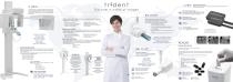
X-View comprises a wide range of digital imaging devices that using state-of-the-art technology produces and manages two and three-dimensional images. X-View is an innovative tool to support doctors making decisions on accurate diagnostics and best therapies. RiX-70 DC High frequency Intraoral X-Ray Unit X-View has multiple fields of application in addition to the applications designed exclusively for Implantology or Endodontics, it helps in the diagnosis of pathologies related to Periodontology, Orthodontics and maxillofacial surgery: Diagnosis of TMJ disorders A detailed evaluation of bone morphology...
Open the catalog to page 2All Trident catalogs and technical brochures
 X-VIEW 2D PAN BASIC
4 Pages
 Brochure I-View
2 Pages
 SOLE CLASS B AUTOCLAVE
2 Pages
 X-View 3D Cone Beam
5 Pages
- Flat panel detector
- Dental radiography system
- Digital dental radiography system
- Intraoral X-ray system
- Waterproof flat panel detector
- Panoramic X-ray system
- Digital panoramic X-ray system
- Digital intraoral X-ray system
- Cephalometric X-ray system
- 70 kV dental radiography
- 60 kV dental radiography
- CR scanner
- Dental CBCT scanner
- Panoramic X-ray system with cephalometric X-ray
- Panoramic X-ray system with CBCT scanner
- Wall-mounted dental radiography system
- Wall-mounted intraoral X-ray system
- 70 kV intraoral X-ray system
- 60 kV intraoral X-ray system
- Dental CR scanner