
- Catalogs
- US Ophthalmic
- Sapphire

Sapphire

Sapphire
The diagnostic platform requires an Intel® Core™ i7 processor, SSD drive, 8 GB RAM, a screen resolution of 1600x900, and two USB ports (one USB 3.0). It operates on Microsoft® Windows® 8 or 10 Professional x64.
The platform supports 5 MP image resolution, multi-shot and video modes, autofocus, manual focus, and variable ISO. It uses infrared and colored LEDs, with a camera sensitive to infrared and equipped with a yellow filter.
This Class I medical instrument, compliant with En. 60601-1, is designed for tear film tests and Meibomian Glands analysis.
- Meibography: Uses infrared light to visualize and diagnose meibomian gland dropout.
- Anterior Segment Imaging: Assesses tear film stability with non-invasive break-up time (NIBUT).
- Tear Meniscus: Evaluates tear volume by examining the tear meniscus.
- 3D Meibography: Provides 3D imaging of Meibomian Glands.
- Auto Interferometry: Evaluates the lipid layer on the tear film.
- Tear Stability Evaluation: Uses Placid disk projection for tear film stability assessment.
The device analyzes tear film composition to identify Dry Eye Disease (DED) types and treatment options.
- Blink Quality: Analyzes blinking efficiency.
- Blepharitis Detection: Visualizes blepharitis and Demodex presence.
- Ocular Redness Classification: Compares conjunctiva images with redness classification sheets.
- Pupillometry: Measures pupil reaction to light.
- White to White Measurement: Evaluates corneal diameter.
Automatically detects and classifies Meibomian Glands using infrared meibography.
Non-invasive measurement of tear meniscus height as an indicator of tear production.
Analyzes blinking quality, crucial for ocular surface health.
Highlights inflammation and detects bacteria, fungi, or viruses causing blepharitis.
Demodex brevis is a mite causing symptoms in large infestations, feeding on sebaceous glands.
Procedure involves patient history, eye structure examination, and tear quality evaluation to diagnose blepharitis types.
Dr. Luca Vigo and Studio Carones provide a comprehensive approach to diagnosing and treating dry eye conditions using Sapphire software.
Includes bulbar redness classification, pupillometry, and white-to-white measurement for understanding Meibomian Gland dysfunction.
Sapphire software supports treatment management and report creation for patient education and follow-up.
Lists Sapphire package contents and provides contact information for US Ophthalmic.
Catalog excerpts

Integrated and easy to use diagnostic platform
Open the catalog to page 1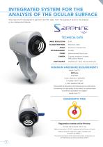
INTEGRATED SYSTEM FOR THE ANALYSIS OF THE OCULAR SURFACE The instrument is designed to perform tear film tests, from the quality of tears to the analysis of the Meibomian Glands. TECHNICAL DATA IMAGE RESOLUTION ACQUISITION MODE Multi shot, video Autofocus, manual focus ISO MANAGEMENT Variable CONES CAMERA LIGHT SOURCE Main cone and Placid cone Colored, sensitive to infrared (NIR), yellow-filtered Infrared LED – Blue, red and white LED MINIMUM HARDWARE REQUIREMENTS Intel® Core™ i7 SSD Drive 8 GB RAM Screen resolution: 1600x900 1 available USB 3.0 port 1 other available USB port Microsoft® Windows®...
Open the catalog to page 2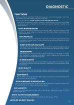
DIAGNOSTIC FUNCTIONS The Device is the new instrument for the individual analysis of tear film that allows to carry out a quick detailed structural research of the tear composition. Research on all the layers (Lipid, Aqueous, Mucin) and Meibomian Glands. Thanks to the Us Ophthalmic Device it is possible to identify the type of Dry Eye Disease (DED) and determine which components can be treated with a specific treatment, in relation to the type of deficiency. AUTO INTERFEROMETRY The Sapphire automatically evaluates the quantity and quality of the lipid component on the tear film. The device highlights...
Open the catalog to page 3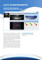
AUTO INTERFEROMETRY COMPLETE MEIBO ANALYSIS: STRUCTURE AND SECRECTION VIEW The evaluation of the lipid layer is part of your overall Dry Eye Assessment. Knowing what is causing Dry Eye will help determine the best treatment option. After your assessment is complete, the Optometrist will discuss your treatment options. Lipid pattern classification, incidence and clinical interpretation is adapted from Guillon & Guillon description incidence (%) with estimated thickness (nm). Observation of blinking frequency and completeness should also be considered - while listening to history and symptoms can...
Open the catalog to page 4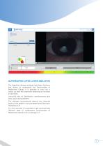
AUTOMATED LIPID LAYER ANALYSIS The Sapphire software analyses lipid layer thickness and allows to understand the functionality of Meibomian Glands. It is possible to carry out a follow up after MG treatment detecting an increase in secretion. Using the new Us Ophthalmic, Interferometry gets easy, quick and automatic. The software automatically detects the coloured lipids on the patient’s eye and determines lipid layer thickness (LLT). In a few seconds it is possible to get automatically relevant data to understand functionality of Meibomian Glands such as average LLT.
Open the catalog to page 5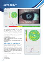
The Device allows to evaluate tear film stability and regularity, using non-invasive break up time measurement (NIBUT). This measures the number of seconds between one complete blinking and the appearance of the first discontinuity in the tear film. With the US Ophthalmic Device, thanks to one single video, the physician can gain lots of information: • Automatic NIBUT • Average of more than one value • Graph to understand the trend of tear film stability during the video • Tear topography that shows all breaking the tear film during time. TEAR STABILITY EVALUATION Through Placid disk projection...
Open the catalog to page 6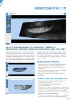
AN OUTSTANDING DIAGNOSTIC EVALUATION IS NEEDED TO DEMONSTRATE THE EFFECTIVENESS OF THE IPL TREATMENT TO PATIENTS The revolutionary introduction of the 3D Meibomian Gland imaging gives two big advantages. Firstly, it enables to confirm the presence of abnormal glands compared to a healthy subject in a 3D view; secondly, it provides a clear image to share with the patients, to help explain the potential reason of their discomfort. Moreover, this new imaging system provides strong evidence to support the choice of a specific therapy (for example IPL treatment) and helps the patient to understand...
Open the catalog to page 7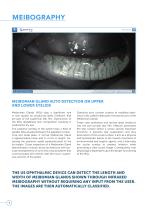
MEIBOMIAN GLAND AUTO DETECTION ON UPPER AND LOWER EYELIDS Meibomian Glands (MGs) play a significant role in tear quality by producing lipids (meibum) that are part of the superficial tear film. Dysfunction of the MGs destabilizes tear composition resulting in evaporative dry eye. The posterior lamella of the eyelid hosts a fleet of parallel MGs situated between the palpebral conjunctiva and tarsal plate. A normal Meibomian Gland is approximately linear and 3–4 mm in length, traversing the posterior eyelid perpendicularly to the lid margin. Closer inspection of a Meibomian Gland demonstrates a...
Open the catalog to page 8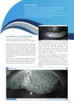
HOW IT WORKS The System automatically analyses the images taken through a sensitive infrared camera (NIR) to locate the Meibomian Glands in a guided way: • An exam valid both for the upper and the lower eyelids; • Automatic percentage of the extension of MGs in the chosen area • Automatic percentage of the Meibomian Gland loss area If the operator prefers, it is also possible to manually compare the images taken with three different related grading scales. AUTOMATIC LID DETECTION To decrease evaluation time, the software automatically detects the lid margin for MG analysis. Meibomian Gland dysfunction...
Open the catalog to page 9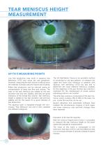
TEAR MENISCUS HEIGHT MEASUREMENT UP TO 5 MEASURING POINTS Low tear production may result in aqueous tear deficiency (ATD) and cause dry eye symptoms. However, measuring the tear volume is difficult since the methods normally used are invasive and irritating. Reflex tear production can be induced, giving an overestimation of basal tear flow and volume. The sizes of the tear meniscus are related to the tear secretion rate and tear stability, and they are good indicators of the overall tear volume. Tear meniscus height is related to the tear secretion rate and tear stability and for this reason...
Open the catalog to page 10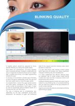
BLINKING QUALITY A healthy person should be expected to show periodic blinking, by closing the eyelids briefly. state of the subjects and the method under which the blink is measured. Most blinks are spontaneous, occurring regularly with no external stimulus. However, a reflex blink can occur in response to external stimuli such as a bright light, a sudden loud noise, or an object approaching towards the eyes. It is also well known that wearing contact lenses (both rigid and soft lenses) can induce significant changes in blinking rate and completeness. A voluntary or forced blink is another type...
Open the catalog to page 11All US Ophthalmic catalogs and technical brochures
 EFC-2600
8 Pages
 LRK-7800
8 Pages
 Auto Ref/Keratometer HRK-1
4 Pages
 BINOCULAR MOBILE REFRACTOMETER
12 Pages
 Surgical Camera System
2 Pages
 Binocular Loupes
3 Pages


Archived catalogs

- Ultrasound system
- Healthcare facility stool
- B/W ultrasound system
- Diode laser
- Medical kit
- Portable ultrasound system
- Tabletop laser
- Examination chair
- Endoscope
- Stool with backrest
- Height-adjustable examination chair
- Electric examination chair
- Emergency kit
- CMOS camera
- Camera with USB port
- Millisecond laser
- Examination chair with legrest
- Class 4 laser
- Surgical loupe
- Examination chair with adjustable backrest