- Catalogs
- Vilber GmbH
- DRI OCT Triton series
DRI OCT Triton series
1 /4Pages

DRI OCT Triton series
1 /4Pages
Catalog excerpts

DRI OCT Triton series Detailed comprehensive reports TOPCOIX YOUR VISION. OUR FOCUS.
Open the catalog to page 1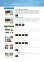
DRI OCT Triton Report view Full comprehensive data analysis GLAUCOMA & MACULA 12 mm x 9 mm 3D Wide scan One rapid scan can cover both the macular and disc areas providing more information for efficient diagnosis. This mode provides macular analysis, thickness map of RNFL, GCL+IPL, RNFL+GCL+IPL and a significance map; all data supporting the diagnosis of macular abnormality and glaucoma. Combination scan This new scan pattern provides both 3D wide scan (12 mm x 9 mm) and Line / 5 Line Cross / Radial scan. Previous OCT models do not offer the option to capture B-scan and 3D images at the same time....
Open the catalog to page 2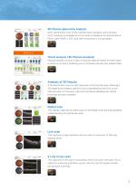
3D Macula glaucoma analysis With vertical box scan of the macular area, Ganglion Cell Complex (GCC) analysis is available and a normative database for Retinal Nerve Fibre Layer (RNFL), GCC and retina thickness is incorporated. Trend analysis (3D Macula analysis) Macular analysis of up to 4 sets of macular data (8 results for both eyes), is shown in a report, enabling you to compare old and new patient data. Analysis of 3D Macula A horizontal box scan can be captured in the macular area, allowing a 3D image to be created; useful for fully understanding the form of the macular area. A thickness...
Open the catalog to page 3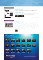
Anterior Line scan Limbus to limbus capture of anterior segment through 16 mm scan. Anterior Radial scan 12 radial scans of the cornea to comprehensively examine the condition of the central cornea. Corneal thickness maps are available. *The anterior module is optional OCT Angiography scan OCT Angiography scans can visualize the retinal microvascular network. It is easy to compare the OCT Angiography image (Superficial, Deep, Outer retina, Choriocapillaris) with the color fundus and B-scan image on a single report. Using IMAGEnet 6, an OCT Angiography image can be overlaid on the color fundus...
Open the catalog to page 4All Vilber GmbH catalogs and technical brochures
 NEWTON 7.0
6 Pages
 SIMPLY E-BOX PRECISE
6 Pages
 E-BOX
E-BOX1 Page
 UV INSTRUMENTS
20 Pages
 Spaide FAF Filters
2 Pages
 INFINITY
1 Page
 FUSION SOLO S
1 Page
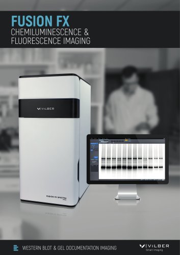
 FUSION FX
1 Page
 FUSION FX SPECTRA
1 Page
Archived catalogs
 NEWTON 7.0 VIVO
1 Page
- Medical lamp
- Veterinary anesthesia system
- Small animal veterinary anesthesia workstation
- Germicidal lamp
- Cell imaging system
- Automatic cell imaging system
- Tabletop veterinary anesthesia system
- Laboratory cell imaging system
- Transilluminator
- Gel documentation system
- Electrophoresis transilluminator
- UV transilluminator
- Gel documentation system with integrated camera
- Fluorescence cell imaging system
- LED transilluminator
- Molecular imaging system
- DNA gel documentation system
- Automatic molecular imaging system
- Molecular biology molecular imaging system
- Fluorescence gel documentation system