optovue iseries
1 /16Pages

optovue iseries
1 /16Pages
Catalog excerpts
optovue iseries Engineered for your OCT success
Open the catalog to page 1
Improving OCT performance with ease When an OCT design puts user experience first, it becomes simple to learn and easy to use. iseries systems are ideal for eye care providers (ECPs) who need advanced OCT technology. Its system software is very intuitive, with helpful helpful graphics and timely prompts that walk you through an exam. Most users are up to speed in a day. Another secret to the iseries’ simplicity is completeness, the capacity for one OCT to deliver the full spectrum of applications from cornea to retina. Add it all up and you arrive at an unparalleled combination of ease and clinical...
Open the catalog to page 3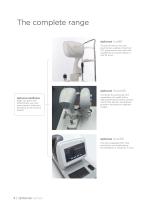
The complete range optovue ivue80 The benchmark for eye care practitioners seeking unmatched OCT performance and value-with capabilities you would expect to cost far more. optovue ifusion80 optovue iwellness helps you grow and differentiate your eye care practice, while also providing a new revenue stream. Combines the advanced OCT capabilities of ivue80 with a high-performance fundus camera icam12 that delivers exceptional posterior and anterior segment images. optovue iscan80 The fully integrated OCT that practically runs itself-setting the standard for simplicity in OCT.
Open the catalog to page 4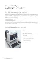
Introducing optovue iscan80® The OCT that practically runs itself Introducing the next-generation iScan - where advanced scanning is realized in a system so user-friendly it even talks to patients. Meet iscan80®, the high-speed 80kHz OCT that sets the standard for efficiency - perfect for ECPs seeking an affordable and versatile OCT system. It’s ideal for practices with limited staff since iscan80 delivers consistent scan acquisition with minimal training - and vocally guides patients through an entire exam in any of 12 languages. The new iscan80 delivers the following advancements: • 80,000...
Open the catalog to page 6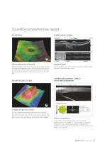
iScan80 comprehensive report 3D RETINA Retinal analysis and 3D imaging Enhanced Depth Retinal imaging capabilities include retina mapping with normative comparison and a 3D retina scan with en face presentation, which enables virtual retinal dissection by displaying three different reference planes: ILM, IPL and RPE. 12mm widefield scan with enhanced depth imaging mode provides high resolution views. 3D OPTIC DISC SCAN ANTERIOR SEGMENT ANGLE WITH MEASUREMENT In-depth 3D optic disc analysis 3D Disc scans are used as the reference to calculate and draw the outline of the optic disc to be used as...
Open the catalog to page 7
Introducing optovue ivue80® HIGH-SPEED 80KHZ OCT + ICAM12 FUNDUS CAMERA OPTION 80,000 A-scans per second - 3x faster than the original iVue OCT improved efficiency and enhanced image quality Simplified scan acquisition real-time en-face imaging displays a 12x9mm view of the retina during acquisition to assist operator in scanning the desired location New reports and wider field of view enhanced capabilities make iVue80 one of the best values in OCT technology today High-resolution fundus and external photography add iCam12 to iVue80 to bring detailed documentation and additional return on your...
Open the catalog to page 8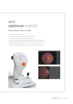
and optovue icam12 NON-MYDRIATIC FUNDUS CAMERA • 45° color and red-free imaging • 12-megapixel camera for high-fidelity color saturation • Multi-visit view provides visit-to-visit comparison • hree-color display offers varying perspectives of the fundus while the T emboss feature creates a 3D-like view for new insights into retinal health • Overlay feature to superimpose OCT images onto the fundus photo • External color photography documents conditions of the ocular surface
Open the catalog to page 9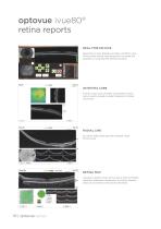
optovue ivue80® retina reports REAL-TIME EN FACE Real-time en face display provides a 12x9mm view of the retina during scan acquisition to assist the operator in scanning the desired location. 3D RETINA CUBE 7x7mm cube scan provides visualization of 201 raster lines to enable in-depth analysis of retinal structures. RADIAL LINE Six 12mm radial lines provide multiple views of the retina. RETINA MAP Visualize a 9x5mm area of the retina with an ETDRS reference database comparison to quickly identify areas of increased or decreased thickness.
Open the catalog to page 10
optovue ivue80® glaucoma reports GLAUCOMA 3D DISC CUBE REPORT 6x6mm cube scan provides visualization of 201 raster lines to enable in-depth analysis of optic disc structures. GANGLION CELL COMPLEX (GCC) ANALYSIS The GCC thickness map allows identification and measurement of ganglion cell loss in glaucoma and Optovue’s exclusive Focal Loss Volume metric (FLV%) is the single best predictor of conversion to glaucoma.1 NERVE FIBER LAYER ANALYSIS The nerve fiber thickness map allows visualization and quantification of RNFL thinning in glaucoma. COMPREHENSIVE REPORTS iVue80’s GCC and RNFL analysis...
Open the catalog to page 11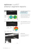
optovue ivue80® anterior segment reports PACHYMETRY AND EPITHELIAL THICKNESS MAPPING (MAPPING ÉPITHÉLIAL AVAILABLE AS OPTION) Visualize and quantify 6mm of epithelial, stromal and total corneal thickness to identify areas of thickening or thinning related to dry eye disease, keratoconus, or previous refractive surgery. The Change Analysis report measures changes in thickness between visits. VAULT MAP REPORT (OPTIONAL) Visualize the fluid reservoir between the lens and cornea for precise scleral lens fitting. ANGLE SCAN Assess angle structure with a quick, non-contact scan and quantify angle parameters...
Open the catalog to page 12
optovue iwellness protocol The iwellness protocol is a valuable assessment tool that can reveal the need for more extensive imaging. It also streamlines the exam process by quickly confirming normal or else aiding in more efficient diagnosis of pathology. In addition, wellness programs improve patient involvement and retention for practice differentiation and growth. iwellness generates a single, comprehensive report to promote better overall eye health. The report includes a 12x9mm structural scan that optimizes metrics on retinal thickness and ganglion cell thickness to the superior/inferior...
Open the catalog to page 13
Technical specifications optovue ivue80 Scanner OCT Image 80,000 A-scans/second Depth Resolution (in tissue) 5.0 μm Transverse Resolution 15 μm (retina) Scan range Depth 2 - 2.3mm (retina) Scan Beam Wavelength 840nm (+/-10nm) OCT Fundus image face FOV Minimum Pupil Diameter External Image (Live IR) FOV Dimensions Dimensions (in.) Weight (lb.) Computer/Networking Specifications Operating System Processor Speed 3.0 GHz; Intel Quad Core (desktop); Core 2 (laptop) Monitor Resolution DICOM compatible Network Bandwidth Computer R
Open the catalog to page 14All Visionix catalogs and technical brochures
 weco e6 s line
12 Pages
 weco e5 s line
8 Pages
 weco e5
12 Pages
 weco e32
8 Pages
 weco e12
6 Pages
 weco e7
6 Pages
 2022 eye refract
16 Pages
 2022 vx40
6 Pages
 vx650
vx65012 Pages
 vx100 serie
12 Pages
 vx65
vx658 Pages
 nexy A.I
8 Pages
 | optovue solix
16 Pages
 Nexus,
4 Pages
 briot scan8
6 Pages
 briot perception2
6 Pages
 briot evolution
8 Pages
 briot emotion2
6 Pages
 briot couture
8 Pages
 briot attitude evolution
8 Pages
 briot attitude
8 Pages
 VX650 Diagnostic
8 Pages
 phoropter vx50 visionix
2 Pages
 VX90 VISIONIX
4 Pages
 Visionix VX110
4 Pages
 WECO E.5
6 Pages
 VX130
VX1306 Pages
 VX118
VX1184 Pages
 VX120
VX1206 Pages
 VX110
VX1104 Pages
 VX40
VX402 Pages
 VX 60
VX 602 Pages
 VX55
VX554 Pages
 VX130+
8 Pages
 VX120+
6 Pages
 VX90
VX904 Pages
 VX36
VX364 Pages
 perception2
6 Pages
 emotion2
6 Pages
 E.32 weco
6 Pages
 E.12 weco
4 Pages
 C.4
C.44 Pages
 Evolution GT
4 Pages
 VX 40
VX 402 Pages
 C.6
C.64 Pages
 Attitude
12 Pages
 Evolution
6 Pages
 PERCEPTION
6 Pages
 E.6
E.66 Pages
 E.5
E.56 Pages
 E.3
E.34 Pages
 WECO E.1
4 Pages
 Eye Refract
8 Pages
 VX 55
VX 554 Pages
 VX 120
6 Pages
 EMOTION
6 Pages
 VX 24
VX 242 Pages
 VX 22
VX 224 Pages
 C.5 The art of blocking
4 Pages
 WECO C.6
6 Pages
 VX BOX II
4 Pages
Archived catalogs
 VX 130
6 Pages
- Visualization software
- Cloud-based software
- Fixed ophthalmic examination
- AI-assisted software
- Artificial intelligence software
- Medical software on site
- Hand-held ophthalmic examination instrument
- Workstation with chair
- Slit lamp
- Table slit lamp
- Ophthalmic workstation
- Compact workstation
- Digital video camera
- Automatic optical lens processing system
- Tonometer
- Retinal camera
- Refractometer ophthalmic examination
- Automatic refractometer
- Keratometer ophthalmic examination
- Ophthalmic software