VX650 Diagnostic
1 /8Pages

VX650 Diagnostic
1 /8Pages
Catalog excerpts

the unique Eye health monitor
Open the catalog to page 1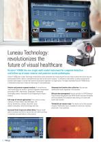
Luneau Technology: revolutionizes the future of visual healthcare Visionix® VX650: the one single multi-modal instrument for complete detection and follow-up of major anterior and posterior ocular pathologies. Visionix® VX650 from Luneau Technology revolutionizes ocular assessment by introducing the first and unique solution that allows eye care professionals (ECPs) to deliver a comprehensive eye exam at the push of a button. It combines an aberrometer, a fundus camera and all essential technologies to monitor both anterior and posterior segments in a single device. The highly automated Visionix®...
Open the catalog to page 2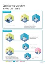
Optimise your work flow on your own terms SYNCHRONOUS Testing, data review and patient consultation happen in the same appointment. Testing is in one location while the data review and patient consultation take place in another location. Same time and location for testing, data review and patient consultation. Patient testing takes place at one time and location while the data review and patient consultation take place at a different time and location. Supervision of eye examination with remote assistance. Secretary completes the patient file. The doctor remotely reviews the data coming from the...
Open the catalog to page 3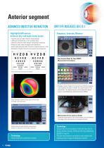
Anterior segment ADVANCED OBJECTIVE REFRACTION Highlights differences between day and night vision needs DRY EYE DISEASES (D.E.D.) Diagnose, Evaluate, Monitor > Objective day and night refraction measurements > 1400 points analysed for a 7-mm diameter pupil > Objective refraction under mesopic and photopic conditions > Measures lower-order and higher-order aberrations > Access visual acuity and quality of vision on a pupil as small as 1.2 mm > Modulation Transfer Function curve analysis and comparison > Non-Invasive Break Up Time (NIBUT) Measurement and analysis Vision quality and Visual acuity...
Open the catalog to page 4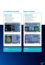
CORNEAL TOPOGRAPHY Diagnose, Evaluate, Monitor Cataracts Diagnose, Evaluate, Monitor Keratoconus > Visualization of lens opacities > Corneal, Internal and wavefront analysis > Internal astigmatism measurement > Kappa angle for IOL centering > Z.4.0 value for aspheric implant > Lens opacity classification (LOC III scale) > Axial, tangential elevation and refraction maps > Keratoconus probability index (KPI) > Keratoconus monitoring > Internal astigmatism measurement > Eccentricity and meridian tables > Corneal aberrometry Retroillumination to examine lens opacities Topography maps and Keratoconus...
Open the catalog to page 5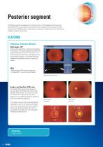
Posterior segment Retinography captures the appearance of a patient’s fundus. The photographs allow the eye care professional (ECP) to study a patient’s retina, detect retinal changes and review a patient’s retinal finding. Visionix® VX650 enables a simple diagnostic procedure to identify patients who need prompt treatment to prevent loss of vision. GLAUCOMA Diagnose, Evaluate, Monitor Irido angle < 20° Angle-closure glaucoma is a medical eye emergency. If the pressure is not reduced quickly, the patient may have permanent vision loss. It is important to note that some patients with narrow angle...
Open the catalog to page 6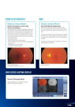
DIABETIC RETINOPATHY Diagnose, Evaluate, Monitor Diagnose, Evaluate, Monitor Diabetic retinopathy can lead to other serious eye conditions: Age-related Macular Degeneration Over time, about half of patients with diabetic retinopathy will develop Diabetic macular edemea (DME). DME happens when blood vessels in the retina leak fluid, causing swelling in the macula (a part of the retina). If a patient has DME, their vision will become blurry because of the extra fluid in their macula. The eye care professional will look at the retina for early signs of the disease, such as: > Leaking blood vessels,...
Open the catalog to page 7
Technical specifications DIMENSIONS / WEIGHT Measurement range Pachymetry, IC (iridocorneal) angle and pupillometry Method Static horizontal scan with the Scheimpflug camera Pachymeter measuring range Pachymetry resolution IC angle measuring range Pupil illumination Retroillumination Corneal topography by specular reflection Calibrated range 7 - 44 mmHg GENERAL Alignment 10.1” (1024 x 600) TFT screen Multi-touch screen Observation area Medical device directive Power mapping and refraction Spherical power range Cylinder power range Measuring area Number of measuring points Acquisition time Number...
Open the catalog to page 8All Visionix catalogs and technical brochures
 weco e6 s line
12 Pages
 weco e5 s line
8 Pages
 weco e5
12 Pages
 weco e32
8 Pages
 weco e12
6 Pages
 weco e7
6 Pages
 2022 eye refract
16 Pages
 2022 vx40
6 Pages
 vx650
vx65012 Pages
 vx100 serie
12 Pages
 vx65
vx658 Pages
 nexy A.I
8 Pages
 | optovue solix
16 Pages
 optovue iseries
16 Pages
 Nexus,
4 Pages
 briot scan8
6 Pages
 briot perception2
6 Pages
 briot evolution
8 Pages
 briot emotion2
6 Pages
 briot couture
8 Pages
 briot attitude evolution
8 Pages
 briot attitude
8 Pages
 phoropter vx50 visionix
2 Pages
 VX90 VISIONIX
4 Pages
 Visionix VX110
4 Pages
 WECO E.5
6 Pages
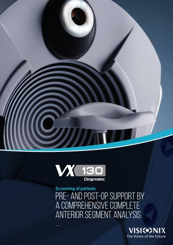
 VX130
VX1306 Pages
 VX118
VX1184 Pages
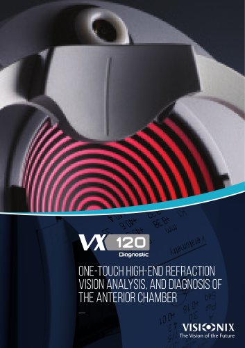
 VX120
VX1206 Pages
 VX110
VX1104 Pages
 VX40
VX402 Pages
 VX 60
VX 602 Pages
 VX55
VX554 Pages
 VX130+
8 Pages
 VX120+
6 Pages
 VX90
VX904 Pages
 VX36
VX364 Pages
 perception2
6 Pages
 emotion2
6 Pages
 E.32 weco
6 Pages
 E.12 weco
4 Pages
 C.4
C.44 Pages
 Evolution GT
4 Pages
 VX 40
VX 402 Pages
 C.6
C.64 Pages
 Attitude
12 Pages
 Evolution
6 Pages
 PERCEPTION
6 Pages
 E.6
E.66 Pages
 E.5
E.56 Pages
 E.3
E.34 Pages
 WECO E.1
4 Pages
 Eye Refract
8 Pages
 VX 55
VX 554 Pages
 VX 120
6 Pages
 EMOTION
6 Pages
 VX 24
VX 242 Pages
 VX 22
VX 224 Pages
 C.5 The art of blocking
4 Pages
 WECO C.6
6 Pages
 VX BOX II
4 Pages
Archived catalogs
 VX 130
6 Pages
- Visualization software
- Cloud-based software
- Fixed ophthalmic examination
- AI-assisted software
- Artificial intelligence software
- Medical software on site
- Hand-held ophthalmic examination instrument
- Workstation with chair
- Slit lamp
- Table slit lamp
- Ophthalmic workstation
- Compact workstation
- Digital video camera
- Automatic optical lens processing system
- Tonometer
- Retinal camera
- Refractometer ophthalmic examination
- Automatic refractometer
- Keratometer ophthalmic examination
- Ophthalmic software