
- Catalogs
- West Medica
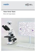
- Vision Hema® Basic Blood cell image analysis

Vision Hema® Basic Blood cell image analysis

Vision Hema® Basic Blood cell image analysis
Digital microscopy integrates microscopes, cameras, software, and computers to manage and analyze microscopy data. It provides qualitative and quantitative results that are otherwise difficult or costly to obtain. West Medica prioritizes innovation in digital microscopy, focusing on hematology, microscopy, cytogenetics, quality control, clinical laboratory, and general medicine.
Vision Hema® Basic Benefits
1. Standardizes analysis results, particularly in blood smear analysis.
2. Documents analysis with storage of images, results, and comments.
3. Facilitates accurate diagnosis through comprehensive data registration.
4. Reduces personnel workload, minimizing fatigue and physical strain.
5. Enhances professional skills and promotes information exchange.
Manual Blood Cell Capture Procedure
The process involves selecting the number of cells, focusing on them, capturing images, and automatic identification and pre-classification of leucocytes.
Autofocus Feature
Ensures automatic lens focusing with a single button press for clear images.
Blood Cell Identification and Classification
White blood cells (WBC) are differentiated into 15 groups, red blood cells (RBC) into 6 groups, and platelets into 3 groups. Laboratory personnel validate and confirm analysis results.
Analysis Results and Interpretation
Includes CBC data, WBC differential, erythrocyte parameters, and platelet analysis. Results are interpreted with notes.
Data Management and Archiving
The Vision Hema® Basic database stores patient records and analysis results, allowing remote access and dynamic assessment. The system automatically saves data, preventing loss.
Reporting and Quality Control
Offers customizable report templates and an atlas for professional development. Quality control assesses diagnostic sensitivity and efficiency.
Specifications
Includes a biological microscope, digital camera, and software for blood cell image capture and analysis. Features include autofocus, pre-classification, and customizable reporting.
Blood Analysis Stages
1. Screening: Use of hematology analyzers.
2. Pre-analytical: Standardized blood smear preparation.
3. Analytical: Blood cell capture and analysis using Vision Hema® Basic.
Contact Information
West Medica, Franz-Siegel-Gasse 1, 2380 Perchtoldsdorf, Austria. Tel: +43 (1) 804 81 84, Email: [email protected].
Catalog excerpts
Vision Hema® Basic Blood cell image analysis
Open the catalog to page 1
Vision HEMAtology West Medica Digital microscopy Digital microscopy is a digital environment for managing and analyzing microscopy data, which is obtained using a microscope, a camera, software and a computer. Digital microscopy allows you to attain qualitative and quantitative results, which are either impossible to receive by other means or cost and time consuming. West Medica feels strongly that innovation is the key to a brighter future and improves the quality of life for everyone. Development of “Vision” solutions for digital microscopy is a top priority and the main focus of West Medica....
Open the catalog to page 2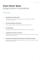
Vision HEMAtology Vision Hemа® Basic Budget solution for laboratories 5 main benefits Standardization of analysis results Vision Hema® Basic system implements quality standards in the most difficult area of hematology — the analysis of a blood smear. Documented analysis of a blood smear Storage of all data, including blood cell images, analysis results, marks and comments. Accurate diagnosis A complete set of data and registration of all objects of interest are a key to accurate diagnosis. A significant load reduction on personnel Working with Vision Hema ® Basic system does not lead to...
Open the catalog to page 3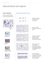
Vision HEMAtology Manual blood cell capture Quick configuration Manual scanning procedure One click on a ‘colour settings’ button to configure the camera and get a high quality digital slide. Move the stage, focusing on the blood cells. Сapture the desired images of blood cells (leucocytes, erythrocytes, platelet). When a leucocyte is in the field of view, the system automatically identifies and hightlights it with a «frame». Select the number of cells required for analysis. Once the desired number of blood cells is collected, the system will start to identify and pre-classify. white balance...
Open the catalog to page 4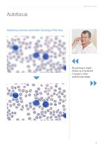
Vision HEMAtology Autofocus Autofocus ensures automatic focusing of the lens By pressing a single button on a keyboard, I can get a clear and focused image
Open the catalog to page 5
Vision HEMAtology Identification, pre-classification and validation of blood cells WBC with differentiation in 15 groups*: RBC with differentiation in 6 groups: 1. Band neutrophiles 2. Segmented neutrophiles 3. Eosinophiles 4. Basophiles 5. Monocytes 6. Blasts 7. Promyelocytes 8. Prolymphocytes 9. Myelocytes 10. Metamyelocytes 11. Lymphocytes 12. Large granular lymphocytes 13. Plasma cells 14. Reactive lymphocytes 15. Unknown Erythrocytes Proerythroblasts Basophilic erythroblasts Polychromatophilic erythroblasts Oxyphilic erythroblasts Erythrocytes with inclusions Platelets with differentiation...
Open the catalog to page 6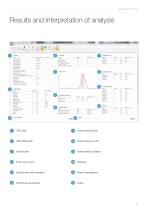
Vision HEMAtology Results and interpretation of analysis Price-Jones curve Erythrocytes with inclusions Result interpretation Erythrocyte parameters
Open the catalog to page 7
VISION HEMATOLOGY Database for archive keeping Data management and archive Vision Hema® Basic database stores all patient records, sample data, analysis results and reports. Information is shown in a form of a patient record with analysis result. Possibility to assess quickly the dynamics in analysis results over a period Digital archive with blood smear samples allows remote viewing of analysis, as well as access to patient's archive. The amount of patient information and analysis results stored is limited only by your disk space. Reliable system The system saves all data automatically, excluding...
Open the catalog to page 8
Vision HEMAtology Report Various features Atlas Develop your professional expertise by creating and maintaining an atlas. Add images with comments for later reference. Quality control Assessment of diagnostic sensitivity, specificity and efficiency. Signals about problems and errors to correct blood smear preparation procedure or service the functional parts of the system. Diagnoses Adjustable reference guide of diagnoses allows to track the formulation, modification or clarification of the diagnosis. Vision system offers report templates for every case From simple analysis on 1/3 of the page...
Open the catalog to page 9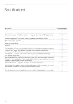
Vision HEMAtology Biological microscope with “infinitive” optics and a stage for 1 slide, Vision CAM® digital camera. Personal computer with Vision Hema® Basic software and a high resolution monitor. Patient and analysis registration. Blood cell image capture. Autofocus. Pre-classification of blood cells, including differentiation of leucocytes, erythrocytes and platelets. “Shortcut” keys, masks, quick preview, color marks and comments for captured cells in the sample. Drag&Drop feature. Complete blood count form. Customizable reference guide to generate reports, following your personal requirements....
Open the catalog to page 10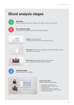
Vision HEMAtology Blood analysis stages 1 Pre-analytical stage Use your hematology analyzer. Samples with «flags» are sent for microscopy. Standardization and automation of blood smear preparation. V-Mixer® hematology mixer. Thorough mixing is achieved by constant spinning. V-Sampler® Instrument for preparation of standardized blood smears. High quality blood smears. V-Chromer® III automated stainer for blood smears. Strict compliance with May-Grünwald Giemsa. Analytical stage Blood cell capture and analysis Vision Hemа® Basic Budget solution for laboratories — — — — — Standardization of analysis...
Open the catalog to page 11
Vision HEMAtology www.vision-hema.com www.wm-vision.com We reserve the right to change specification without notice. Product images are shown for reference only and final product may differ Official distributor
Open the catalog to page 12All West Medica catalogs and technical brochures
 Vision Hema® Network
1 Page
 Vision Hema® Integro
4 Pages
 V-Counter® PC-analyzer
4 Pages
 Vision Hema® Remote
4 Pages
 V-Counter® PC Analyzer
4 Pages
 Vision Master catalogue 2014
62 Pages


Archived catalogs

- Cell imaging system
- Automatic cell imaging system
- Hospital management system
- Laboratory cell imaging system
- Fluorescence cell imaging system
- Laboratory management system
- Diagnostic cell imaging system
- Molecular biology cell imaging system
- Phase contrast cell imaging system
- Bright field cell imaging system
- Blood cell cell imaging system
- Hematology cell imaging system
- Clinical cell imaging system
- Veterinary laboratory cell imaging system
- Karyotyping cell imaging system
- FISH cell imaging system