- Catalogs
- West Medica
- Vision Hema® ? Blood cell image analysis systems
Vision Hema® ? Blood cell image analysis systems

Vision Hema® ? Blood cell image analysis systems
Vision Hema® is a digital microscopy system by West Medica, designed to automate blood cell analysis for various laboratory applications, including hematology and oncology. It aims to enhance diagnostic accuracy and laboratory productivity.
- Automation and Standardization: Automates blood smear analysis, including scanning and pre-classification of blood cells.
- Improved Productivity: Increases diagnostic accuracy and reduces manual workload.
- Ergonomics and Efficiency: Minimizes physical strain and optimizes workflow.
- Professional Development: Supports continuous learning and expertise sharing.
- Network Capabilities: Facilitates data exchange and remote consultations.
- Vision Hema® Assist: Cost-effective for small labs, focusing on cell identification.
- Vision Hema® Pro: Suitable for small to medium labs with enhanced scanning capabilities.
- Vision Hema® Ultimate: For large labs, featuring advanced automation.
- Workflow Optimization: Streamlines processes and reduces manual tasks.
- Quality Assurance: Provides reliable analysis results.
- Time Efficiency: Automates routine tasks, saving time.
- Collaboration: Enables real-time information sharing.
- Automatic Scanning: Classifies blood cells into groups like WBC, RBC, and platelets.
- Flexible Interface: User-friendly with Drag&Drop and shortcuts.
- Data Management: Comprehensive database for analysis results.
- Remote Access: Supports remote connections for data sharing.
The process involves selecting cell groups, choosing analysis methods, classifying objects, and generating reports, ensuring accurate analysis.
Offers customizable templates including patient information and CBC data, tailored to specific needs.
Stores patient records and analysis results, supporting remote access for efficient information sharing.
Integrates traditional microscopy with automated scanning, maintaining classical features while enhancing efficiency.
Facilitates data sharing and collaboration across locations, supporting video conferencing.
Aids in education by providing tools for reviewing blood cell images and facilitating discussions.
Includes leucocyte distribution detection, quality control, and adaptive scanning, supporting virtual sample creation.
Emphasizes standardizing blood smear preparation for accurate analysis, including automated mixing and staining.
Outlines equipment options like motorized microscopes and automated slide stainers, highlighting their benefits.
Catalog excerpts

Vision Hema® Blood cell image analysis systems
Open the catalog to page 1
Vision HEMAtology West Medica Digital microscopy Digital microscopy is a digital environment for managing and analyzing microscopy data, which is obtained using a microscope, a camera, software and a computer. Digital microscopy allows you to attain qualitative and quantitative results, which are either impossible to receive by other means or cost and time consuming. West Medica feels strongly that innovation is the key to a brighter future and improves the quality of life for everyone. Development of “Vision” solutions for digital microscopy is a top priority and the main focus of West Medica....
Open the catalog to page 2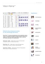
Vision HEMAtology Vision Hema® Applications clinical laboratory blood transfusion stations Efficient and professional solution for the hematology laboratory! Vision Hema® is a blood cell identification and pre-classification system. Its aim is to automate and simplify a complex procedure of blood smear analysis. You work with a system where priority is given to efficiency and workflow optimization and standardization. research Vision Hema® Assist — digital system for blood cell image analysis. Cost-effective solution for small-sized laboratories Vision Hema® Pro — digital system for blood cell...
Open the catalog to page 3
Vision HEMAtology Workflow automation and standardization Improved quality and increased productivity Efficient use of time and correct ergonomics Standardize your work and analysis results. All stages of blood smear analysis are optimized: automatic scanning, identification and pre-classification of blood cells, validation of analysis results, data archive and report generation. Provide a reliable diagnosis. Analysis is accurate and objective thanks to automatic recognition and preclassification of blood cells. Microscopic blood smear analysis documentation and specialized software provide greater...
Open the catalog to page 4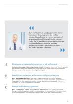
Vision HEMAtology Care and attention to qualified personnel are very important in the management of a working process. It’s much easier to view an automatically generated gallery of blood cells on the computer rather than to spend hours looking into microscope eyepieces. Reduction in routine workload on qualified personnel significantly facilitates the work of the entire laboratory Continuous professional development of lab technicians Benefit from knowledge and experience of your colleagues Internet and network capabilities Increase the knowledge and perfect professional skills. By working with...
Open the catalog to page 5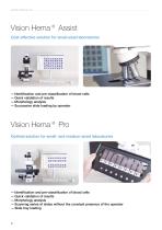
Vision HEMAtology Vision Hema® Assist Cost-effective solution for small-sized laboratories — Identification and pre-classification of blood cells — Quick validation of results — Morphology analysis — Successive slide loading by operator Vision Hema® Pro Optimal solution for small- and medium-sized laboratories — Identification and pre-classification of blood cells — Quick validation of results — Morphology analysis — Scanning series of slides without the constant presence of the operator — Slide tray loading
Open the catalog to page 6
Vision HEMAtology Vision Hema® Ultimate Effective solution for large-sized laboratories — Identification and pre-classification of blood cells — Quick validation of results and morphology analysis — Scanning series of slides without the constant presence of the operator — Random Access and STAT testing — Continuous loading mode — Automatic oil dispenser — Automatic slide loader
Open the catalog to page 7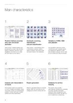
Vision HEMAtology Flexible interface provides easy and convenient operation Automatic scanning, identification and pre-classification Vision Hema® flexible interface can be learned by a user in few minutes. Variety of features («shortcut» keys, Drag&Drop, etc) allow you to keep the work style you are used to. Vision Hema® automatically scans a microscopic sample, collects data for analysis, sorts blood cells and saves results in the database. The specialist is free from routine monotonous work. Analysis results are displayed in galleries of cells and arranged in groups. All you need to do is...
Open the catalog to page 8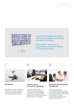
Vision HEMAtology Automation through Vision Hema® improves the quality and increases the speed of analysis Vision Hema® allows you to save time and resources, wasted on routine microscopy Remote access and network capabilities Education and professional development Comfortable work with a high quality microscope. Automatic approach to blood smear analysis, retaining the traditional classic microscopy. Connecting several working places to a remote server allows the organization of an information network for the medical institution. Organize video conferences and share analysis results with colleagues....
Open the catalog to page 9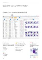
VISION HEMATOLOGY Easy and convenient operation It only takes a click to see patient records and analysis results Analysis status Analysis status is used for convenient management of a laboratory workflow Visual signals when analysis results Show group of blood cell group Leave your comments and marks right on the blood cell image
Open the catalog to page 10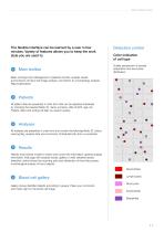
Vision HEMAtology The flexible interface can be learned by a user in few minutes. Variety of features allows you to keep the work style you are used to. Main toolbar Detection control Color indication of cell type Quality assessment of sample preparation and leucocytes distribution. Basic working tools. Management of patients records, analysis results and functions of blood cell image analysis. Connection to a hematology analyzer. Report generation. All patient data are presented in chart form that can be adjusted individually by choosing the required fields: ID, name, surname, date of birth,...
Open the catalog to page 11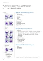
VISION HEMATOLOGY _ Automatic scanning, identification * EaiyJ reutroohiles • Scgmtnttt r.«vt'Op>ilrl Largt orimitir lyinornxytn tryitvod CfOi ' Basophilic efyth'oblast * OxyptHlic efythrobUs* • Erythrocytes with exclusion WBC with differentiation in 15 groups" 12. Large granular lymphocytes RBC with differentiation in 6 groups — Identification by size (normocytes, microcytes, macrocytes, megalocytes) — Identification by color (normochromic, polychromatophilic, hypochromic, — Identification by shape (microspherocytes, target cells, ovalocytes, stomotocytes, sickle cells, schistocytes, acanthocytes,...
Open the catalog to page 12All West Medica catalogs and technical brochures
 Vision Hema® Network
1 Page
 Vision Hema® Integro
4 Pages
 V-Counter® PC-analyzer
4 Pages
 Vision Hema® Remote
4 Pages
 V-Counter® PC Analyzer
4 Pages
 Vision Master catalogue 2014
62 Pages


Archived catalogs

- Cell imaging system
- Automatic cell imaging system
- Hospital management system
- Laboratory cell imaging system
- Fluorescence cell imaging system
- Laboratory management system
- Diagnostic cell imaging system
- Molecular biology cell imaging system
- Phase contrast cell imaging system
- Bright field cell imaging system
- Blood cell cell imaging system
- Clinical cell imaging system
- Hematology cell imaging system
- Veterinary laboratory cell imaging system
- Karyotyping cell imaging system
- FISH cell imaging system