- Catalogs
- ZAP Surgical Systems Inc
- Stereotactic radiotherapy for metastatic brain tumors

Stereotactic radiotherapy for metastatic brain tumors
Stereotactic radiotherapy for metastatic brain tumors
Brain metastases affect 20% to 40% of cancer patients, with treatment options including surgery, targeted therapy, immunotherapy, and radiation therapy. This study evaluates radiotherapy techniques for metastatic brain tumors, focusing on NC-VMAT, C-VMAT, Helical TomoTherapy, CyberKnife, Gamma Knife, and ZAP-X.
The study involved 12 patients treated with CyberKnife for a single brain tumor. Treatment plans were created for each device, divided into stereotactic radiosurgery (SRS) and stereotactic radiotherapy (SRT) groups. Parameters evaluated included Gradient Index (GI), Paddick Conformity Index (CI), and treatment time (t-time).
In the SRS group, ZAP-X and Gamma Knife showed superior CI and GI for small tumors, while NC-VMAT and C-VMAT had shorter treatment times. In the SRT group, ZAP-X and Gamma Knife maintained superior CI and GI, with NC-VMAT and C-VMAT again having shorter treatment times. ZAP-X, CyberKnife, and Gamma Knife had longer treatment times regardless of tumor volume.
ZAP-X is a new gyroscopic radiosurgery device offering advantages in CI and GI similar to Gamma Knife, especially for small tumors. The study emphasizes selecting appropriate radiotherapy techniques based on tumor size and treatment time considerations.
ZAP-X shows promise in treating small metastatic brain tumors with favorable dosimetry parameters, though treatment time remains a consideration. Further studies are needed to explore its full potential compared to established techniques like Gamma Knife and CyberKnife.
The study evaluates dose indicators V12 Gy and V24 Gy for normal brain tissue. In the SRS group, all techniques maintained V12 Gy below 10.0 cc, while in the SRT group, V24 Gy exceeded 16.8 cc. The mean values of GI and CI were generally better for Gamma Knife compared to other techniques.
For the SRS group, C-VMAT had the shortest treatment time, followed by NC-VMAT and Helical TomoTherapy. In the SRT group, similar trends were observed. ZAP-X, CyberKnife, and Gamma Knife had longer treatment times, influenced by factors such as imaging time and beam repositioning.
Limitations include the small sample size and variability in tumor volume, which may affect results. The study also acknowledges potential differences in results due to the use of different planning systems and equipment.
ZAP-X shows relatively superior CI and GI for small tumors, similar to Gamma Knife. However, for larger tumors, differences between techniques are less pronounced. VMAT techniques offer shorter treatment times, making them suitable when time efficiency is a priority.
Catalog excerpts
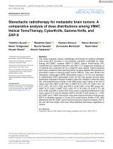
RESEARCH ARTICLE Stereotactic radiotherapy for metastatic brain tumors: A comparative analysis of dose distributions among VMAT, Helical TomoTherapy, CyberKnife, Gamma Knife, and ZAP-X Toshihiro Suzuki1 Naoto Yanagisawa4 Hiroshi Onishi2 Ryutaro Nomura3 Hikaru Nemoto2 Zennosuke Mochizuki1 Naoki Sano2 1 CyberKnife Center, Kasugai General Rehabilitation Hospital, Yamanashi, Japan 2 Department of Radiology, University of Yamanashi, Yamanashi, Japan 3 Department of Neurosurgery, Kamiyacho Neurosurgical Clinic, Tokyo, Japan 4 Division of Radiation Oncology, Aizawa Comprehensive Cancer Center, Aizawa Hospital, Nagano, Japan 5 Department of Neurosurgery, Kasugai General Rehabilitation Hospital, Yamanashi, Japan Correspondence Masahide Saito, Department of Radiology, University of Yamanashi, 1110 Shimokato, Chuo-city, Yamanashi, 409–3898, Japan. Email: [email protected] Abstract This study evaluates various radiotherapy techniques for treating metastatic brain tumor (BT), focusing on non-coplanar volumetric modulated arc radiotherapy (NC-VMAT), coplanar VMAT (C-VMAT), Helical TomoTherapy (HT), CyberKnife (CK), Gamma Knife (GK), and ZAP-X. CT images and structures of 12 patients who underwent CK for a single BT were utilized. Twelve treatment plans were created for each planning device. All plans adopted the approach of prescription doses to planning target volume D99.5%. They were divided into stereotactic radiosurgery (SRS) (prescription dose; 21–23 Gy) and stereotactic radiotherapy (SRT) (prescription dose; 30–36.5 Gy) groups and the same parameters evaluated included Gradient Index (GI), Paddick Conformity Index (CI), and treatment time (t-time). In the SRS group, mean values of GI and CI values were: NC-VMAT (4.28, 0.60), C-VMAT (5.61, 0.44), HT (4.68, 0.42), CK (4.31, 0.61), GK (2.81, 0.82), and ZAP-X (2.99, 0.80). In the SRT group: NCVMAT (3.27, 0.84), C-VMAT (3.81, 0.82), HT (3.76, 0.65), CK (2.98, 0.77), GK (2.61, 0.90), and ZAP-X (2.80, 0.84). There were no significant differences in the mean values of CI and GI between ZAP-X and GK in both groups (p > 0.05). NC-VMAT and C-VMAT had shorter t-time than other techniques in both groups. ZAP-X is relatively superior in CI and GI for small tumors, similar to GK, while differences with NC-VMAT and CK diminish as tumor volume increases. ZAP-X, CK, and GK have longer t-time than other treatment techniques, regardless of volume. KEYWORDS CyberKnife, Gamma Knife, Helical TomoTherapy, metastatic brain tumor, VMAT, ZAP-X This is an open access article under the terms of the Creative Commons Attribution License, which permits use, distribution and reproduction in any medium, provided the original work is properly cited. © 2025 The Author(s). Journal of Applied Clinical Medical Physics published by Wiley Periodicals, LLC on behalf of The American Association of Physicists in Medicine. J Appl Clin Med Phys. 2025;e70046. https://doi.org/10.1002/acm2.70046
Open the catalog to page 1
Brain metastases are reported to manifest in approximately 20% to 40% of the entire cohort of cancer patients.1,2 Treatment options include surgical intervention, targeted therapy, immunotherapy, and radiation therapy. Furthermore, there are cases where these are combined.3 Presently, within the domain of radiotherapy, an array of techniques is available, including volumetric modulated arc radiotherapy (VMAT), stereotactic radiosurgery (SRS), and stereotactic radiotherapy (SRT).4 Treatment devices encompass a range of instruments, including linear accelerator (LINAC), Helical TomoTherapy (HT,...
Open the catalog to page 2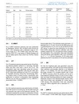
JOURNAL OF APPLIED CLINICAL TABLE 1 Characteristics of the 12 patients. Number of Prescription Target size Target Patient Age Sex Primary lesion Fraction dose (Gy) (cc) localization R frontal R parietal L frontal R occipital R temporal R cerebellum Colon cancer Rectum cancer Melanoma Melanoma Breast cancer Lung cancer L frontal R occipital L frontal R cerebellum R parietal L frontal Note: Patients 1 to 6 were considered the stereotactic radiosurgery group, patients 7 to 12 were designated as the stereotactic radiotherapy group. Prescription doses and number of fractions were calculated using...
Open the catalog to page 3
The multiple isocenters and collimators were selected to align with the shape and size of the PTV. For some cases, forward planning as well as GK was used to shape the 50% isodose line according to the shape of the tumor, while inverse planning was used to calculate the other cases. The collimator sizes used were 1–3 selected from 4, 5, 7.5, 12.5, and 15 mm for the SRS group and 3–5 selected from 7.5, 10, 12.5, 15, 20, and 25 mm for the SRT group. In other cases, inverse planning was performed after forward planning. This involves conducting forward planning with the isocenter placement intended...
Open the catalog to page 4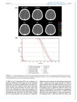
NC-VMAT Normal brain tissue C-VMAT Normal brain tissue HT Normal brain tissue CK Normal brain tissue GK Normal brain tissue ZAP-X Normal brain tissue NC-VMAT PTV C-VMAT PTV HT PTV CK PTV GK PTV ZAP-X PTV FIGURE 1 The dose distribution (a) and DVH (b) for representative cases (patient 3: PTV 0.3 cc, Prescription dose 22 Gy) of each of the six planning techniques in the SRS group. (green line: Region surrounded by a dose of 12 Gy) *CK utilizes the Monte Carlo algorithm, resulting in dose distribution plots in the air. DVH, dose-volume histogram. C-VMAT, and HT. Comparing ZAP-X as a reference, no...
Open the catalog to page 5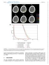
JOURNAL OF APPLIED CLINICAL (Gy) NC-VMAT Noma I brain tissue-NC-VMAT PTV C-VMAT Normal brain tissue C-VMAT PTV HT Normal brain tissue -HT PTV CK Normal brain tissue -CK PTV GK Normal brain tissue -GKPTV ZAP-X Normal brain tissue -ZAP-X PTV FIGURE 2 The dose distribution (a) and DVH (b) for representative cases (patient 9: PTV 24.0 cc, Prescription dose 36.5 Gy) of each of the six planning techniques in the SRT group. (green line: Region surrounded by a dose of 24 Gy) *CK utilizes the Monte Carlo algorithm, resulting in dose distribution plots in the air. DVH, dose-volume histogram. and CK. Comparing...
Open the catalog to page 6