- Catalogs
- Zimmer Biomet
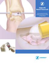
- DeNovo® NT Natural Tissue Graft Arthroscopically-Assisted Surgical Technique for Ankle Cartilage Repair
DeNovo® NT Natural Tissue Graft Arthroscopically-Assisted Surgical Technique for Ankle Cartilage Repair

DeNovo® NT Natural Tissue Graft Arthroscopically-Assisted Surgical Technique for Ankle Cartilage Repair
DeNovo NT Natural Tissue Graft is a human tissue allograft used for repairing articular cartilage lesions in the ankle. It consists of juvenile hyaline cartilage pieces with viable chondrocytes and is secured using fibrin sealant, eliminating the need for periosteal flap harvesting and suturing. It is suitable for lesions larger than 1cm2 or as a secondary treatment for failed previous repairs.
Pre-operative imaging such as MRI or CT scans is recommended to assess lesion size and location. The graft is typically used for lesions greater than 1cm2. One pack of tissue graft is recommended per 2.5cm2 of lesion, with a fill ratio of at least 50%.
Address ligament laxity and consider ligament reconstruction to ensure stable joint mechanics. Correct axial deformity with osteotomy if necessary.
Joint distraction methods such as traction or weighted ankle wraps are suggested to facilitate lesion visualization and access.
Autologous fibrin is recommended for graft fixation. Ensure fibrin is ready before lesion filling, and have multiple delivery tips available due to potential obstructions.
- Arthroscopy: Use a 2.7mm or 1.9mm arthroscope for lesion inspection. Perform tibial plafondoplasty if needed for better visualization.
- Prepare the Cartilage Lesion: Curette the lesion to a stable border, removing affected cartilage to the subchondral plate. Ensure at least 50-60% of the lesion is bordered by native cartilage.
- Create a Viable Bone Bed: If the subchondral bone is sclerotic, perform procedures like curettage or microfracture to achieve a spongy base.
- Dry the Lesion and Create an Extended Portal Incision: Ensure a dry environment for graft delivery. Extend the portal incision for better access.
- Prepare DeNovo NT Graft: Introduce graft packages aseptically, aspirate preservation medium, and prepare for delivery.
- Load DeNovo NT Graft into a Delivery Cannula: Use a Freer elevator or spatula to load cartilage pieces into a cannula.
- Cover Lesion Base with Fibrin: Apply fibrin to the lesion base, ensuring it does not adhere to opposing surfaces.
- Deliver DeNovo NT Graft: Extrude cartilage pieces into the lesion before fibrin cures, ensuring at least 50% coverage.
- Set and Test: Allow graft to cure, apply additional fibrin if needed, and ensure the graft does not impinge in the joint.
Remove debris, perform standard closure, and apply a splint with the ankle in a neutral position.
- Weeks 0-6: Non-weight bearing with a posterior plaster splint, transitioning to a removable boot brace.
- Weeks 6-12: Begin weight-bearing as tolerated, increase range of motion exercises, and wean from the brace.
- Weeks 12-52: Gradually increase activity, progressing to running and loading activities.
Catalog excerpts
DeNovo NT Natural Tissue Graft ® Arthroscopically-Assisted Surgical Technique for Ankle Cartilage Repair
Open the catalog to page 1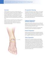
Arthroscopically-Assisted Surgical Technique for Ankle Cartilage Repair Pre-operative Planning DeNovo NT Natural Tissue Graft is an off-the-shelf human tissue allograft, consisting of juvenile hyaline cartilage pieces with viable chondrocytes, and is intended for the repair of articular cartilage lesions in a single-stage procedure. The DeNovo NT Graft surgical technique eliminates the need for harvesting and suturing of a periosteal flap, as it employs fibrin sealant to secure the particulated tissue pieces into the lesion. Pre-operative planning may include an MRI or CT scan to better estimate...
Open the catalog to page 2
Arthroscopically-Assisted Surgical Technique for Ankle Cartilage Repair Arthroscopy Perform a diagnostic arthroscopy to visualize the ankle lesion using a 2.7mm or 1.9mm arthroscope to inspect the lesion (Fig. 1). Removal of the distal tibial cortical bone anterior to the cartilage surface of the tibia (tibial plafondoplasty) may be performed as needed to obtain adequate visualization of posterior lesions. Fig. 1 External view of the arthroscope and internal arthroscopic view of lesion. Prepare the Cartilage Lesion Curette the lesion to a stable border (margin), removing the affected cartilage...
Open the catalog to page 3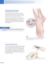
Arthroscopically-Assisted Surgical Technique for Ankle Cartilage Repair Dry the Lesion and Create an “Extended Portal” Incision It is vital to deliver the DeNovo NT Graft and fibrin sealant into a dry environment (whether under a “dry scope” or open arthrotomy procedure) to prevent the graft tissue from being washed away and to allow the fibrin sealant to cure. Turn off and remove the arthroscopic inflow. Apply traction as required to provide enough working joint space, and extend the portal incision by approximately 1-2cm . Clean and dry the lesion with an appropriate curved suction tube, gauze...
Open the catalog to page 4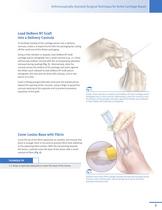
Arthroscopically-Assisted Surgical Technique for Ankle Cartilage Repair Load DeNovo NT Graft into a Delivery Cannula To facilitate loading of the cartilage pieces into a delivery cannula, create a scooped funnel with the packaging by cutting off the small end of the blister packaging. Using a Freer elevator or spatula, load DeNovo NT Graft cartilage pieces retrograde into a small cannula (e.g., a 1.9mm arthroscopy outflow cannula with the accompanying obturator removed during loading) (Fig. 5). Alternatively, slide the cannula across the bottom of the package and press against the blister pack...
Open the catalog to page 5
Arthroscopically-Assisted Surgical Technique for Ankle Cartilage Repair Deliver DeNovo NT Graft After about 1-2 minutes and before the fibrin fully cures, introduce the loaded cannula, and use the obturator to extrude the DeNovo NT Graft cartilage pieces into the lesion and on top of the fibrin (Fig. 7). Ensure that at least 50% of the lesion area is covered with uniformly distributed tissue pieces. In areas that are not fully shouldered, it is recommended that a gap of approximately 1mm be left between the tissue pieces and the edge of the lesion to minimize the risk of graft delamination. While...
Open the catalog to page 6
Arthroscopically-Assisted Surgical Technique for Ankle Cartilage Repair Final Inspection and Close Check for any extraneous debris or dislodged cartilage pieces and remove with forceps or gentle suction. Perform a standard closure with suture, and apply a splint with the ankle in a neutral position (Fig. 10). Post-operative Care General post-operative guidelines include: Final tissue/ fibrin construct • Non-weight bearing for 6 weeks – Weeks 0 -2: Posterior plaster splint with the ankle in neutral position is used until sutures are removed – Weeks 2-6: A generic removable boot brace is used....
Open the catalog to page 7
This documentation is intended exclusively for physicians and is not intended for laypersons. Information on the products and procedures contained in this document is of a general nature and does not represent and does not constitute medical advice or recommendations. Because this information does not purport to constitute any diagnostic or therapeutic statement with regard to any individual medical case, each patient must be examined and advised individually, and this document does not replace the need for such examination and/or advice in whole or in part. Please refer to the package inserts...
Open the catalog to page 8All Zimmer Biomet catalogs and technical brochures
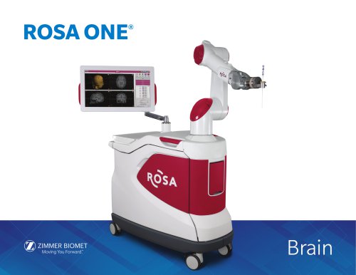 ROSA ONE® Brain System
7 Pages
 MODULAR FEMORAL Revision System
14 Pages

Archived catalogs
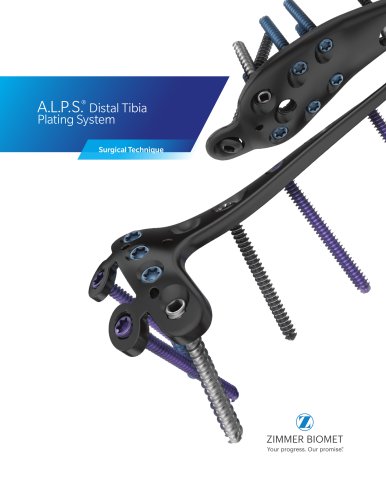 A.L.P.S.®
44 Pages
 Constrained Posterior Stabilized
12 Pages
 Persona PERSONALIZED KNEE
7 Pages
 Avenir® Femoral Hip System
12 Pages
 The CLS® Spotorno® Stem
16 Pages
 Alloclassic®Zweymüller®Stem
12 Pages
 ®Zimmer® Segmental System
6 Pages
 Zimmer Natural Nail System
8 Pages
 modern-cementing-technique
16 Pages
 CoAxial Spray Kit
8 Pages
 Biologics
24 Pages
 PowerPump DP System
2 Pages
 Sidus
Sidus40 Pages
 Ankle Fix System 4.0
8 Pages
 Anatomical Shoulder System
6 Pages
 NexGen® RH Knee
8 Pages
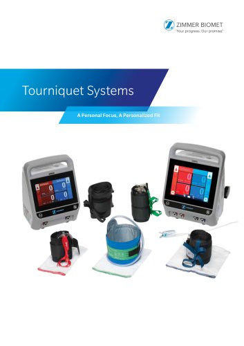
 tourniquet-systems-brochure
8 Pages
 Persona-Partial
12 Pages
 Fitmore Hip Stems
6 Pages
 NexGen High-Flex Implant
8 Pages
 Trabecular Metal ™Glenoid
4 Pages
 Anatomical Shoulder ™ System
6 Pages
 Zimmer ® PSI Shoulder
6 Pages
 Zimmer personna
12 Pages
 ZImmer iASSIST
44 Pages
 Fitmore ® Hip Stem
24 Pages
 Persona Knee
6 Pages
 Trauma Solutions
10 Pages
 Colagen Repair Patch
2 Pages
 Fitmore® Hip Stem
6 Pages
 Zimmer® Segmental System
6 Pages


- Detection kit
- ZIMMER BIOMET bone plate
- ZIMMER BIOMET compression plate
- ZIMMER BIOMET metallic compression plate
- Immunoassay detection kit
- ZIMMER BIOMET locking compression plate
- Infectious disease detection kit
- Titanium compression plate
- Surgical system
- Distal compression plate
- Chromatographic immunoassay test kit
- Cutting electrosurgical system
- Cassette detection kit
- Coagulation electrosurgical unit
- Agitator
- ELISA detection kit
- Proximal compression plate
- Benchtop agitator
- Arthrodesis nail
- Forearm compression plate