- Catalogs
- Zirkonzahn
- Brochure Detection Eye
Brochure Detection Eye
1 /20Pages

Brochure Detection Eye
1 /20Pages
Catalog excerpts

Intraoral scanner for the digital workflow
Open the catalog to page 1
Enrico Steger, Zirkonzahn founder and CEO 2 Julian Steger, Zirkonzahn CEO
Open the catalog to page 2
OUR PRECISION AND SUPPORT RIGHT FROM THE BEGINNING OF THE TREATMENT With our Detection Eye intraoral scanner, we have added to our product package the initial building block for the complete digitisation of Zirkonzahn’s workflow! With each new product, we always remain true to our work philosophy: with Detection Eye, the interaction between clinic, laboratory and patient is brought to the next level, thanks to the innovative software functions and the scanner’s complete integration into our workflow and data management system. Our intraoral scanner will transform chair time into a comfortable...
Open the catalog to page 3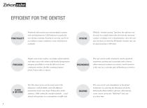
Notebook with touchscreen and touchpad; scanner with anti-fog function, LED indicator to guide the user during scanning; heating of scan tips can be set according to usage conditions; extra-oral function available Simple scan of any surface, to produce photorealistic, real-time scans with colours and detailed preparation margins; possibility to scan the full arch in one continuous motion; flexible scanning distance (from 5 mm to direct contact) The 3D colour scans can be easily sent to the laboratory with flexibility and with different automation levels (via cloud, Zirkonzahn.Archiv software,...
Open the catalog to page 4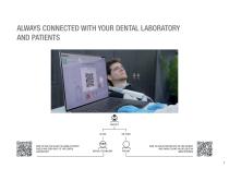
ALWAYS CONNECTED WITH YOUR DENTAL LABORATORY AND PATIENTS HOW TO USE THE CLOUD TO HANDLE PATIENT CASES AND SEND DATA TO THE DENTAL LABORATORY DENTAL TECHNICIAN HOW TO USE DETECTION EYE ON THE PATIENT AND SHARE SCANS VIA QR CODE ON SMARTPHONES
Open the catalog to page 5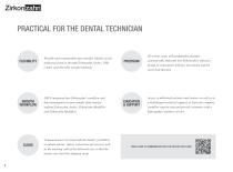
PRACTICAL FOR THE DENTAL TECHNICIAN Flexible and customisable data transfer, ideally via the dedicated cloud or through Zirkonzahn.Archiv, USB, e-mail, and other file transfer methods 3D colour scans with preparation margins automatically imported into Zirkonzahn’s software; design of restorations with jaw movements and the extra-oral function SMOOTH WORKFLOW 100 % integrated into Zirkonzahn’s workflow and data management system (simple data transfer to/from Zirkonzahn.Archiv, Zirkonzahn.Modellier and Zirkonzahn.Modifier) EDUCATION & SUPPORT Access to dedicated webinars and courses as well as...
Open the catalog to page 6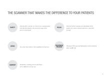
THE SCANNER THAT MAKES THE DIFFERENCE TO YOUR PATIENTS Autoclavable scan tips, no risk of cross-contamination and infection thanks to the practical usage of the device by the dentist Less chair time thanks to the simplified work process TREATMENT PREVIEW Selection of the scanning tone depending on the patient, for a more relaxed experience, especially for kids Sharing of 3D scans and information on the treatment plan via QR code Powderless scanning process and choice of two different scan tip sizes
Open the catalog to page 7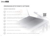
ZIRKONZAHN.DETECTIONEYE SOFTWARE OVERVIEW OF CHARACTERISTICS Open output formats: export as STL, OBJ and PLY Intuitive and user-friendly interface for an efficient work Pre-design functions: pre-visualisation of full scans, alignment of scans, tooth marking, bite and undercut control, automatic margin line detection Pre-settings for intra- or extra-oral usage Scanning of the full arch in one continuous motion Data can be saved on the cloud with no extra costs and with illimited space A special function facilitates scanning of metal parts Artificial intelligence to identify and remove automatically...
Open the catalog to page 8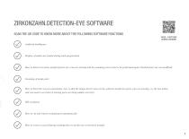
ZIRKONZAHN.DETECTION-EYE SOFTWARE SCAN THE QR CODE TO KNOW MORE ABOUT THE FOLLOWING SOFTWARE FUNCTIONS VIDEO – FUNCTIONS DURING SCANNING Artificial intelligence Display of undercuts (useful during teeth preparation) How to block scan areas (useful if parts of a scan are missing and the scanning process has to be performed again; blocked parts are not modified) Scanning of metal parts How to block the scan in a particular view so that the image doesn’t move in the software (useful if a parts scan are missing, e.g. the last molar, and you need to see better if missing parts are being added correctly)...
Open the catalog to page 9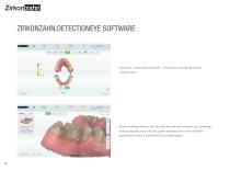
ZIRKONZAHN.DETECTIONEYE SOFTWARE Occlusion control and correction – Green parts indicate the correct contact points. Green markings indicate that the area has not been scanned; grey markings indicate that the scan is in low quality and that extra scans should be performed to reach a maximum level of information.
Open the catalog to page 10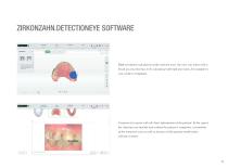
ZIRKONZAHN.DETECTIONEYE SOFTWARE High-resolution calculation of the selected area: the user can select with a brush an area that has to be calculated with high precision, for example in case of dies or implants. Creation of a report with all clinic information of the patient. In the report, the clinician can include notes about the patient’s symptoms, screenshots of the intraoral scan as well as pictures of the patient mouth taken with the scanner.
Open the catalog to page 11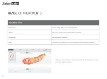
RANGE OF TREATMENTS TREATMENT TYPE Restoration Crown, bridge, inlay, veneer and antagonist Full crown, small to large-span bridges on implants Oral exam, caries filling, root canal treatments and periodontal treatments Abutments and scanbodies can be scanned directly into the patient’s mouth to transfer implant positions.
Open the catalog to page 12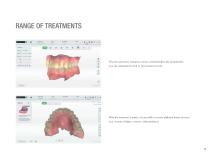
The pre-operatory situation can be scanned before die preparation (e.g. the anatomical tooth or provisional crown). With the intraoral scanner it is possible to create different kinds of cases (e.g. crowns, bridges, veneers, inlays/onlays).
Open the catalog to page 13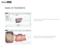
The scanner permits the digitisation of every kind of orthodontic treatment (e.g. aligners, bite splints). The patient’s mouth can be scanned during the first appointment, to take note of the patient’s clinical information overtime.
Open the catalog to page 14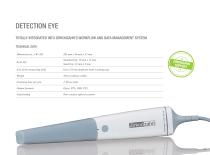
DETECTION EYE TOTALLY INTEGRATED INTO ZIRKONZAHN’S WORKFLOW AND DATA MANAGEMENT SYSTEM TECHNICAL DATA Dimensions (L x W x H) Scan tips Size of the measuring field Up to 22 mm depth for both scanning tips Output formats Non-contact optical scanner
Open the catalog to page 15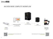
AN EVEN MORE COMPLETE WORKFLOW PRINTING DESIGNING AND POSITIONING NEW! Zirkonzahn.Modifier and Zirkonzahn.Slicer software CURING NEW! L300 Post-Curing Lamp PLASTER-FREE ARTICULATION NEW! Detection Eye intraoral scanner MORE INFORMATION ABOUT THE P4000 PRINTING SYSTEM READ THE BROCHURE NEW! P4000 Printer and Printer Resin Waterbased Beige MORE INFORMATION ABOUT THE P4000 PRINTING SYSTEM VISIT THE WEBPAGE
Open the catalog to page 16All Zirkonzahn catalogs and technical brochures
 Case gallery Prettau® Skin®
4 Pages
 Case gallery Prettau® Skin®
4 Pages
 Insert ICE Plus
4 Pages
 Insert X-Ray Sphere
4 Pages
 Insert Cocronit Superior
4 Pages
 Insert Clara Zanini
4 Pages
 Case gallery Prettau® Skin®
4 Pages
 Insert Zirkonofen Turbo
4 Pages
 Insert anti-snoring device
4 Pages
 Insert Gingiva-Composites
4 Pages
 Brochure Fresco Ceramics
52 Pages
 Brochure PlaneSystem®
28 Pages
 Insert Zirkonofen 600/V4
4 Pages
 Fresco Ceramics Application
6 Pages
 Prettau® 3 Dispersive®
4 Pages
 Brochure JawAligner
28 Pages
 Brochure PlaneSystem®
24 Pages
 Flyer PlaneAnalyser II
1 Page
 Flyer Head Tracker
1 Page
 Brochure CAD/CAM System
88 Pages
 Insert M6 Blank Changer
5 Pages
 Case gallery Bartplatte
4 Pages
 Brochure Zirkonzahn thinks big
12 Pages
 Brochure Shade Guide
52 Pages
 Insert Prettau® Line
4 Pages
 Case gallery Fresco Ceramics
4 Pages
 Brochure Zirkonofen Turbo
36 Pages
 Case gallery Prettau® 2
4 Pages
 Implant-supported components
100 Pages
 Case gallery Raw-Abutment®
4 Pages
 Insert Software
6 Pages
 Case gallery Bite Splint
4 Pages
 Brochure My Laboratory
32 Pages
 Brochure Zirkonzahn
68 Pages
 Brochure Military School
32 Pages
 Brochure Sintering Furnaces
28 Pages
 Brochure Patients
24 Pages
 Brochure Material diversity
116 Pages


Archived catalogs


- ZIRC dental material
- ZIRC dental restoration material
- Implant abutment
- Titanium implant abutment
- Straight implant abutment
- Dental surgery instrument kit
- Dental burr
- ZIRC resin dental material
- Visualization software
- Internal implant abutment
- ZIRC dental prosthesis dental material
- ZIRC heating oven
- Modeling dental material
- ZIRC dental crown material
- ZIRC biocompatible dental material
- Diamond burr
- Angled implant abutment
- Dental bridge material
- Benchtop oven
- Dental laboratory dental material