
Excertos do catálogo

Innovation with Integrity Label-Free Molecular Imaging • Discover, localize and quantify biochemical changes and molecular markers Mass Spectrometry
Abrir o catálogo na página 1
Bruker, the global leader in MALDI-MS technology, is also your partner for label-free molecular MALDI Imaging MALDI is the most robust ion desorption technology for analyzing many different classes of analyte molecules from many types of samples. When MALDI spectra are collected in a spatially orientated pattern, each spectrum represents a molecular fingerprint of a specific and unique region of the sample being analyzed. Any detected ion can be projected into a 2 dimensional map of sample location and intensity – an ion image. From a single dataset, hundreds to thousands of unique,...
Abrir o catálogo na página 2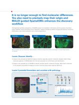
It is no longer enough to find molecular differences. You also need to precisely map their origin and MALDI guided SpatialOMx enhances the discovery workflow Homogenized samples analyzed by LC-MS/MS present no localization information and the process may dilute highly localized compounds below limit of detection. Analyze samples by MALDI Imaging and capture compound distributions, even when localized to very small compartments. MALDI Imaging can simultaneously map hundreds of different compounds within a small region or across large cohorts of samples. Locate, Discover, Identify ... •...
Abrir o catálogo na página 3
N-Glycan maps show glycolylations specific to colorectal cancer PNGase F treatment liberates N-linked glycans • Visualize patterns of glycosylation • Identify N-glycans using MS/MS • Correlate glycosylation patters to pathology Trypsin cleaves intractable proteins into detectable peptides • Tryptic peptides reveal distribution of larger proteins in fresh frozen and FFPE tissues • Identify protein from peptide MS/MS • Access insoluble proteins colorectal cancer Richard R. Drake Professor and Director of MUSC Proteomics Center, Medical University of South Carolina, Charleston, SC "We have...
Abrir o catálogo na página 4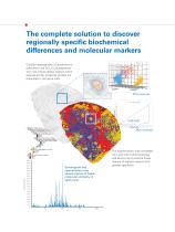
Cellular heterogeneity of lymphoma is reflected in the SCiLS Lab segmentation map where cellular regions which express similar molecular profiles are I presented in the same color. ✓ ' The segmentation map correlates very well with tissue pathology and allows one to analyze those regions of highest interest with greater specificity. Absolute Intensity shows regions of higher molecular similarity in same color.
Abrir o catálogo na página 5
Systems for highest sample throughput Flex MALDI-TOF systems offer the most versatile analytical range and speed while producing hundreds of ion images per sample. autoflex maX: outstanding entry-level MALDI system for Imaging and much more. TOF/TOF is available. Layer of purkinje cells visible ultrafleXtreme: High performance TOF/TOF imaging system 3 phospholipids mapped from rat brain section ultrafleXtreme: high performance TOF/TOF imaging system rapifleX: novel laser optics designed specifically for highest performance and highest speed for MALDI Imaging. TOF/TOF is available.
Abrir o catálogo na página 6
Systems for highest information content scimaX and 2xR MRMS systems offer the highest mass resolving power of any commercial MALDI system and are the ultimate systems for imaging drugs, metabolites and lipids. timsTOF fleX combines the best proteomics platform and MALDI Imaging. Discover MALDI guided SpatialOMx for the most complete discovery workflow
Abrir o catálogo na página 7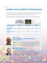
Localize and quantify for drug discovery Make critical decisions earlier: The low operational cost of MALDI Imaging allows it to be deployed very early in the discovery pipeline to help make decisions faster and bridges the gap between traditional LC-MS and Quantitative Whole Body Autoradiography (QWBA) for distribution and metabolism studies. Whether part of dosing and PK studies or for ADME/Tox studies, MALDI Imaging offers high sensitivity, multiple target localization, and simultaneous discovery of toxicity markers – all integrated with histology. MALDI Imaging Spatial distribution...
Abrir o catálogo na página 8
Visually explore biochemical pathways Control Dopamine GABA * Images courtesy of Prof. Per Andrén, Uppsala Univ. Prof. Per Andrén Uppsala University, Sweden “ ass spectrometry imaging enables us to simultaneously map and quantitate M multiple neurotransmitters, their precursors and metabolites directly in tissue sections. That is, almost the complete dopaminergic and serotonergic neurotransmitter networks.
Abrir o catálogo na página 9
Acquire • Method driven image acquisition • Define measurement areas from microscopic image • Image all of the sample or just regions of interest Visualize • Multiple ion overlays • ROI segmentation • Save targeted ions for instant visualization or explore interactively Analyze • SCiLS Lab Core: auto-segmentation and co-localization • SCiLS Cloud: integrates remote teams • SCiLS Lab Pro: full analyses of large sample cohorts • SCiLS Lab Premium 3D: full analyses of 2- and 3-dimensional images
Abrir o catálogo na página 10
Add molecular specificity to other imaging techniques Fluorescence Look beyond optical morphology. Import many other imaging modalities to examine your MSI data in the most relevant context and boost confidence in your analyses. Histology* MALDI Imaging* Whole Tissue Atlas Reference IHC Bruker pioneered the integration of MALDI Imaging data with other imaging modalities to provide you with the most biologically relevant information. Bruker’s imaging software allows co-registration and overlays of multiple images with full user control. * Image courtesy of Dr. Jeff Spraggins, Mass...
Abrir o catálogo na página 11
Contact your local Bruker representative to learn more about our MALDI products and how they can expand your research into the visual world of MALDI Imaging For the highest spatial resolution Bruker offers the industry leading TM Sprayer from HTX Imaging For research use only. Not for use in clinical diagnostic procedures. Bruker Daltonik GmbH Bremen · Germany Phone +49 (0)421-2205-0 ms.sales.bdal@bruker.com - www.bruker.com Scan the QR-Code for more Details Bruker Daltonics is continually improving its products and reserves the right to change specifications without notice. © BDAL 05-2018,...
Abrir o catálogo na página 12Todos os catálogos e folhetos técnicos Bruker Daltonics Inc.
-
 MALDI Biotyper® CA System
12 Páginas
-
 IntelliSlidesTM
2 Páginas
-
 CMC-assist
2 Páginas
-
 Toxtyper
8 Páginas
-
 Product Overview
16 Páginas
-
 maXis II for Biopharma Analysis
8 Páginas
-
 spotOn™
4 Páginas
-
 Metabolomics
8 Páginas
-
 BioPharma Compass® 2.0
8 Páginas
-
 TASQ Software
4 Páginas
-
 timsTOF™
8 Páginas
-
 TargetScreener
6 Páginas
-
 timsTOF Pro
6 Páginas
-
 Quant Proteomics
8 Páginas
-
 MBT Galaxy RUO
4 Páginas
-
 MBT Pilot RUO
4 Páginas
-
 MBT Filamentous Fungi Library
6 Páginas
-
 MBT SMART IVD
4 Páginas
-
 MBT Consumables RUO
4 Páginas
-
 MBT Sepsityper
6 Páginas
-
 MBT BTS US
2 Páginas
-
 MBT Disposable Targets US
2 Páginas
-
 MBT Pharma
8 Páginas
-
 MALDI Imaging
16 Páginas
-
 rapifleX™ MALDI Tissuetyper™
8 Páginas
-
 PesticideScreener 2.0
8 Páginas
-
 MBT Mycobacteria Library
4 Páginas
-
 MALDI Biotyper Clinical IVD
6 Páginas
-
 MALDI Biotyper CA System
12 Páginas
-
 Bruker ToxScreener
6 Páginas
-
 ProteinScape
8 Páginas
-
 maxis II
12 Páginas
-
 EVOQ
EVOQ6 Páginas
-
 impact II
12 Páginas
-
 micrOTOF II
8 Páginas
-
 ImagePrep
4 Páginas
-
 DE-tector
6 Páginas
-
 pTD
pTD2 Páginas
-
 RAID AFM
4 Páginas
-
 RAID XP
4 Páginas
-
 RAID S2
6 Páginas
-
 RAID M100
6 Páginas
-
 MM2
MM26 Páginas
-
 SVG 2 and Probes
6 Páginas
-
 µRAID
µRAID4 Páginas
-
 VeroTect
4 Páginas
-
 SIGIS II
2 Páginas
-
 solarix XR
12 Páginas
-
 The new autoflex speed
10 Páginas
-
 Product Overview
20 Páginas
-
 impact HD
12 Páginas
-
 Radiation Backpack Sentry
4 Páginas
-
 Toxtyper
6 Páginas
-
 micrOTOF-Q III
6 Páginas
-
 #701434 (05-2012) The new ultrafleXtreme
16 Páginas
-
 Brochure autoflex-speed 05-2010 (270289)
10 Páginas
-
 Brochure microflex (Brochure)
6 Páginas


Catálogos arquivados
-
 EVOQ - 2014
6 Páginas