Excertos do catálogo

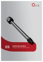
EPIPHYSIS SCREW emergency team for broken bones®
Abrir o catálogo na página 1
• crew material: TiAl6V4 ELI S • Easy removal of the implant after fracture healing • Increased fatigue strenth of the implants • Decreased risk of infection and allergy • annulated Spongiosa Tension C Screw with constant 10 mm thread • ore diameter 5 mm C • uter diameter 6.5 mm O • ength: 50 - 120 mm in 5 mm L steps • annulation: 3.5 mm for Ø 3.2 C mm Guide Wire • elfdrilling & selfcutting S EPIPHYSIS SCREW emergency team for broken bones® Indications • he indication for the use of a T transcutaneous screw holds for all acute, acute to chronic and chronic loosening of the epiphysis •...
Abrir o catálogo na página 2
back-tapping flank large countersink head for easy removableness EPIPHYSIS SCREW
Abrir o catálogo na página 3
Slipped Capital Femoral Epiphysis [SCFE] ECF is a disease of young people and occurs round about sexual maturity. Slippage of the head of the femur occurs on the growth groove. This is really misnamed since it is not the femoral head that moves but rather the metaphysis of the neck of the femur that slips forwards and upwards while the head of the femur is held in the acetabulum by the ligamentum capitis femoris (Fig.1). The problem of this disease is the occurrence of complications such as avascular necrosis or chondrolysis of the head of the femur. Each of these complications can lead to...
Abrir o catálogo na página 4
Preparation of Operation INSTRUMENTS REQUIRED: Extension table and two image intensifiers. EPIPHYSIS SCREW
Abrir o catálogo na página 5
EPIPHYSIS SCREW emergency team for broken bones® The patient is placed on the extension table and two image intensifiers are arranged in such a way that the proximal end of the femur can be represented in two planes. The image intensifiers have to be placed in such a way that the X-ray tubes can be positioned respectively above (A-P level) and between the legs (sagittal level) in order to protect the operating team from X-rays as best as possible. The patient is placed on the extension table with carefully internally rotated leg (neutralising the femoral torsion). Care must be taken not to...
Abrir o catálogo na página 6
EPIPHYSIS SCREW Surgical Technique First step: In the A-P plane, a free Kirschner’s wire is placed centrally over the course of the neck of the femur while being checked with the image converter and fastened to the skin using adhesive strip. This wire determines the middle of the neck of the femur in the A-P plane and thus all possible entry points of the cannulated screw.
Abrir o catálogo na página 7
Second step: Under observation from the image intensifiers in the lateral plane, the point at the centre of the neck of the femur in the A-P plane (where the Kirschner’s wire was attached earlier) is determined to allow an additional central attachment to the epiphysis of the head of the femur in the sagittal plane. The greater the slippage of the head of the femur, the further ventral is the point of entry, and thus the steeper the Kirschner’s wire must be in the lateral plane. EPIPHYSIS SCREW emergency team for broken bones® Insertion of the Kirschner’s wire under observation from the...
Abrir o catálogo na página 8
Third step: Before the measuring device is placed in position, the incision lateral to the guide wire is made larger. Fourth step: Using the measuring device, the length of the appropriate hollow screw is determined. It is necessary, however, to take the distance from the joint into account and therefore to choose a screw some 0.5 cm longer since the Kirschner’s wire stops about 1 cm in front of the joint cavity. EPIPHYSIS SCREW
Abrir o catálogo na página 9
Fifth step: Screw in the hollow screw above the guide wire under observation from the image intensifiers in both planes. Care must be taken not to perforate the joint. EPIPHYSIS SCREW emergency team for broken bones®
Abrir o catálogo na página 10
Sixth and last step: Screw out the guide wire, release the leg, and check mobility using the image intensifiers. Care must be taken not to perforate the joint! EPIPHYSIS SCREW It is important not to countersink the bolt head into the bone, otherwise the easy percutaneous removeableness will not be able. Because of the short screw threat you can use the screw also as a „dynamic“ one. This means that you should retighten the screw due to the growth. For that the head of the screw must stick out from the bone 0.5 – 1 cm (fig.).
Abrir o catálogo na página 11
EPIPHYSIS SCREW emergency team for broken bones® Attaching the screw on the other side The same procedure as in the steps of the operation, except that the guide wire is inserted parallel to the Kirschner’s wire fastened to the skin distal to the tuberculum inominatum.
Abrir o catálogo na página 12
Removing the screw Removal of the epiphysis screw is carried out after closure of the growth groove and checked using the image intensifiers, and is carried out percutaneously. EPIPHYSIS SCREW A guide wire starting from the scar of the screw attachment is introduced into the funnel shaped screw head of the epiphysis screw and the latter removed by means of the T-handle along with the guide wire.
Abrir o catálogo na página 13
Implants & Instruments EPIPHYSIS SCREW emergency team for broken bones® Epiphysis Screw, D=6.5mm, L=50mm, 10mm Thread Epiphysis Screw, D=6.5mm, L=55mm, 10mm Thread Epiphysis Screw, D=6.5mm, L=60mm, 10mm Thread Epiphysis Screw, D=6.5mm, L=65mm, 10mm Thread Epiphysis Screw, D=6.5mm, L=70mm, 10mm Thread Epiphysis Screw, D=6.5mm, L=75mm, 10mm Thread Epiphysis Screw, D=6.5mm, L=80mm, 10mm Thread Epiphysis Screw, D=6.5mm, L=85mm, 10mm Thread Epiphysis Screw, D=6.5mm, L=90mm, 10mm Thread Epiphysis Screw, D=6.5mm, L=95mm, 10mm Thread Epiphysis Screw, D=6.5mm, L=100mm, 10mm Thread Epiphysis Screw,...
Abrir o catálogo na página 14
EPIPHYSIS SCREW Socket Wrench, WS 10, L=250mm, Epiph. Screw Guide Wire, Steel, D=3.2mm, L=260 mm, TR, w. Thrd. Depth Gauge 3.2mm, L=260mm, Epiphys. Wire Sterilization Tray, Epiphysis Screw
Abrir o catálogo na página 15
Chemical process - anodization in a strong alkaline solution * Type III anodization Dotize Type II anodization Different colors Film become an interstitial part of the titanium Implant surface remains sensitive to: Chipping Peeling Discoloration No visible cosmetical effect Anodization Type II leads to following benefits * • • • • • • • • Oxygen and silicon absorbing conversion layer Decrease in protein adsorption Closing of micro pores and micro cracks Reduced risk of inflammation and allergy Hardened titanium surface Reduced tendency of cold welding of titanium implants Increased fatigue...
Abrir o catálogo na página 16Todos os catálogos e folhetos técnicos I.T.S.
-
ufs
1 Páginas
-
DHL
2 Páginas
-
ITS
2 Páginas
-
DHL - Distal Humeral Locking Plates
20 Páginas
-
PHL
24 Páginas
-
ACLS
20 Páginas
-
CFN
32 Páginas
-
OLS
24 Páginas
-
PHLs
20 Páginas
-
CTN - Cannulated Tibia Nail
28 Páginas
-
UOL - Ulna Osteotomy Locking Plate
32 Páginas
-
SR Sacral Rods
20 Páginas
-
HCS
24 Páginas
-
TOS Twist-Off Screw
20 Páginas
-
TLS
20 Páginas
-
PRS-RX
32 Páginas
-
HLS
20 Páginas
-
PLS - Pilon Locking Plates System
24 Páginas
-
ES
20 Páginas
-
SR
20 Páginas
-
FL
24 Páginas
-
PL - Pilon Locking Plate small
12 Páginas
-
PRS - Pelvic Reconstruction System
28 Páginas
-
PRL - PROlock Radius Locking Plate
20 Páginas
-
OHL - Olecranon Hook Locking Plate
24 Páginas
-
OL - Olecranon Locking Plate
24 Páginas
-
PHL - Proximal Humeral Locking Plate
28 Páginas
-
CAS
40 Páginas
-
FCN
20 Páginas
-
HOL
24 Páginas
-
FLS
24 Páginas
-
PFL
20 Páginas
-
DTL
24 Páginas
-
HTO
24 Páginas
-
PTL
32 Páginas
-
DFL
32 Páginas
-
SCL
32 Páginas
-
SLS
24 Páginas
-
CAL
20 Páginas
-
DUL
24 Páginas
-
CLS
28 Páginas


