 Website:
Leica Microsystems
Grupo: Danaher
Excertos do catálogo

ARveo 8 The infinite possibilities of digital neurosurgery start here
Abrir o catálogo na página 1
ARveo 8, the digital visualization microscope for neurosurgery from Leica Microsystems, evolves with you into the digital future enabling your entire neurosurgical team to benefit from a new level of enhanced AR visualization, efficiency and accessibility for more informed and precise neurosurgeries. ARveo 8 unlocks the door to the digital future. The possibilities are infinite.
Abrir o catálogo na página 2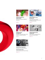
Enhanced visualization for greater precision GLOW AR technology and GLOW800 AR fluorescence Enhanced efficiency across the entire team ARveo 8 graphical user interface, visualization in the OR and beyond with HD 3D viewing and recording Enhanced to unlock the digital future EnhancePath, image guided surgery, robotics integration, endoscope compatibility Enhanced ergonomics make workflows flow ARveo 8 design for comfortable working postures and smooth workflows Enhanced optical image quality FusionOptics and innovative illumination Enhancing patient safety Optimal illumination and fall-back...
Abrir o catálogo na página 3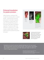
Enhanced visualization for greater precision ARveo 8 enhances visualization. With its ultra-fast processing, latency is reduced by 44%—synchronizing the visual and tactile faster for more precise surgeries.1 The integration of world-renown Leica optics into the GLOW AR ecosystem is at the heart of the ARveo 8 visualization capabilities. They help you obtain more information, so that you can, e.g., safely navigate a complex tangle of abnormal arteries and veins and preserve key nerves and vessels. Anatomical detail Aneurysm viewed in white light High contrast Aneurysm viewed with ICG and NIR...
Abrir o catálogo na página 4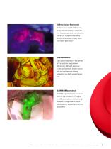
FL400 oncological fluorescence The fluorescence module FL400 is used during open neurosurgery in conjunction with the active substance 5 aminolevulinic acid (5-ALA). It supports resection by allowing differentiation of tumor tissue from healthy brain tissue.2 FL560 fluorescence FL560 allows observation of fluorophores with an excitation range between ~460 nm and ~500 nm. It allows you to view non-fluorescent tissue in natural color and simultaneously observe fluorescence in a bright yellowish-green color. GLOW800 AR fluorescence GLOW800 augmented reality fluorescence takes the high contrast...
Abrir o catálogo na página 5
Enhanced efficiency across the entire team ARveo 8 enhances efficiency. When every second counts in a surgery, anything that slows it down is a barrier to the best possible outcome. ARveo 8 supports a more collaborative workflow for the entire surgical team. Its new GUI strips away interface complexity, keeping only what’s essential for a logically clear path to easy setup and immediate adjustments, at any given point in time. Its 4K external display and multiple display modalities give the full team a better picture and aid in teaching and patient documentation. Right before your eyes One...
Abrir o catálogo na página 6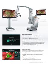
Optional 55-inch 4K 3D cart-mounted monitor 3 for flexible positioning 2-in-1 image display system: The monitor can shows the graphical user interface as well as the microscope image Intuitive graphical user interface The ARveo 8 graphical user interface is designed to be self-explanatory for all members of the OR team. > Assign different roles with different levels of user rights > Be sure user settings cannot accidentally be changed thanks to password protection > Be confident patient and user data are secure thanks to increased cybersecurity > Operate the microscope easily thanks to...
Abrir o catálogo na página 7
Enhanced to unlock the digital future ARveo 8 is ready to fulfill future needs. You can add new technologies as well as AR applications that will impact you, your entire surgical team, and your patients. We call this concept EnhancePath, our promise that ARveo 8 evolves with you into the digital future.
Abrir o catálogo na página 8
Visualization of eloquent fiber tract information Image shows cranial neuronavigation software from BrainLab © KARL STORZ SE & Co. KG, Germany The ability to combine preoperative images with intraoperative imaging can be decisive during procedures. With ARveo 8 you can use image guided surgery systems to overlay your microscope view with additional imaging information, complement it with endoscope imaging as well as use robotic control to keep a specific point of interest in focus. Align and view with ease Navigation-controlled robotics Support your intraoperative assessment with flexible...
Abrir o catálogo na página 9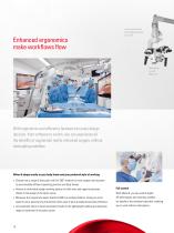
Enhanced ergonomics make workflows flow Long overhead reach for flexible positioning in your OR More space to work (600 mm) With ergonomics and efficiency factored into every design decision, from software to switch, you can experience all the benefits of augmented reality-enhanced surgery, without interrupting workflow. ARveo 8 adapts easily to your body frame and your preferred style of working > Choose from a range of binoculars with full 360°-rotation for main surgeon and assistant to accommodate different operating positions and body frames > Achieve a comfortable upright working...
Abrir o catálogo na página 10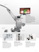
Large overhead clearance Made to withstand The premium overhead stand from our partner Mitaka was designed and built for intensive, flexible, and extremely reliable performance in the OR. Based on aerospace technology, it has a robust, full-metal construction with long reach and a spacesaving compact footprint. Effortless operation via handle or wireless footswitch Compact footprint frees up OR space Smooth balancing Easy maneuvering Save time by pushing a button once for AutoBalancing or twice to balance all six axes. To rebalance the system intraoperatively, even through a sterile drape,...
Abrir o catálogo na página 11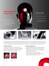
Enhanced optical image quality FusionOptics Technology 1. Two separate optical paths 2. One path provides great depth of field 3. The other path provides high resolution Enjoy the best of two worlds for a significantly expanded area in full focus and less refocusing. Magnification multiplier for 40% boost 4. T he brain effortlessly merges the images into a single, optimal spatial view SpeedSpot for fast focusing Fine focus for rear assistant For too long, surgeons had to compromise between high resolution and greater depth of field – no more! FusionOptics generates two separate beam paths...
Abrir o catálogo na página 12Todos os catálogos e folhetos técnicos Leica Microsystems
-
M844 F40/F20
16 Páginas
-
EnFocus
12 Páginas
-
M822 F40 / F20
12 Páginas
-
M320 for ENT
12 Páginas
-
M320 Dental Brochure
12 Páginas
-
Emspira 3
4 Páginas
-
Exalta
2 Páginas
-
FLEXACAM C1
4 Páginas
-
EM KMR3
8 Páginas
-
EM UC7
16 Páginas
-
EM TRIM2
8 Páginas
-
EM RAPID
8 Páginas
-
EM ICE
12 Páginas
-
EM TXP
10 Páginas
-
EM RES102
12 Páginas
-
EM TIC 3X
16 Páginas
-
TL4000 BFDF
16 Páginas
-
F12 I floor stand
6 Páginas
-
XL Stand
4 Páginas
-
TL3000 Ergo & TL5000 Ergo
4 Páginas
-
KL300 LED
8 Páginas
-
LED1000
16 Páginas
-
LED3000 BLI
20 Páginas
-
LED5000 NVI
20 Páginas
-
LED3000 NVI
20 Páginas
-
LED3000 DI
20 Páginas
-
LED5000 HDI
20 Páginas
-
LED5000 CXI
20 Páginas
-
LED2500
8 Páginas
-
LED5000 MCI
20 Páginas
-
LED3000 MCI
20 Páginas
-
LED5000 SLI
20 Páginas
-
LED3000 SLI
20 Páginas
-
LED2000
8 Páginas
-
LED5000 RL
20 Páginas
-
LED3000 RL
20 Páginas
-
MZ10 F
4 Páginas
-
M165 FC
16 Páginas
-
M205 FCA, M205 FA
16 Páginas
-
M125 C, M165 C, M205 C, M205 A
12 Páginas
-
A60 F, A60 S
16 Páginas
-
M50, M60, M80
12 Páginas
-
DVM6
16 Páginas
-
HCS A
20 Páginas
-
TCS SPE
20 Páginas
-
DFC450 C
6 Páginas
-
DFC295
6 Páginas
-
MC170 HD
6 Páginas
-
DFC3000 G
6 Páginas
-
DMC4500
4 Páginas
-
ICC50 W, ICC50 E
6 Páginas
-
DFC7000 T, DFC7000 GT
4 Páginas
-
DFC9000
2 Páginas
-
IC90 E
6 Páginas
-
DMC6200
8 Páginas
-
DMC5400
8 Páginas
-
SFL7000
4 Páginas
-
EL6000
4 Páginas
-
SFL100
4 Páginas
-
SFL4000
4 Páginas
-
DMi8 S Platform
2 Páginas
-
THUNDER Imager Live Cell
2 Páginas
-
DMi8 M / C / A
12 Páginas
-
DM IL LED
12 Páginas
-
DMi1
6 Páginas
-
DM3 XL
7 Páginas
-
FS M
4 Páginas
-
FS C
4 Páginas
-
FS CB
4 Páginas
-
DM3000, DM3000 LED
16 Páginas
-
DM750 M
12 Páginas
-
DM750
12 Páginas
-
DM500
12 Páginas
-
DM300
8 Páginas
-
DM12000 M
8 Páginas
-
DM8000 M
8 Páginas
-
DM1750 M
12 Páginas
-
DM4 M, DM6 M
12 Páginas
-
DM4 P, DM2700 P, DM750 P
12 Páginas
-
DM2000, DM2000 LED
16 Páginas
-
DM1000
16 Páginas
-
DCM8
16 Páginas
-
DM1000 LED
16 Páginas
-
DM2500
16 Páginas
-
DM4 B & DM6 B
16 Páginas
-
DM6 M LIBS
2 Páginas
-
S9 Series
12 Páginas
-
Z6 APO
16 Páginas
-
Z16 APO
16 Páginas
-
Leica M530 OHX for ENT
4 Páginas
-
M620 F20
8 Páginas
-
M220 F12
8 Páginas
-
2D and 3D IOL guidance systems
8 Páginas
-
Leica Application Suite X
4 Páginas
-
Proveo 8
16 Páginas
-
M525 F20
12 Páginas
-
EnVisu Leica Handheld OCT
8 Páginas
-
PROvido
8 Páginas
-
Leica M530 OHX
16 Páginas
-
Leica TCS SP8 STED 3X
24 Páginas
-
Leica TCS SP8 Objective
24 Páginas
-
Leica AOBS
16 Páginas
-
Leica DMC2900
6 Páginas
-
Leica DMshare
2 Páginas
-
Leica_DM4000-6000-BrochureTechnical
10 Páginas
-
Leica_DMshare_ICC50-Flyer_en
2 Páginas
-
Leica_DMshare_IC80_HD-Flyer_en
2 Páginas
-
Leica_DMshare_EZ4_HD-Flyer_en
2 Páginas
-
Leica_DMshare_EC3-Flyer_en
2 Páginas
-
Leica_SL801-Flyer
2 Páginas
-
Leica_SCN400-Flyer_Clinical
2 Páginas
-
DM2700 M
12 Páginas
-
Leica_SR_GSD_Technical-Brochure
8 Páginas
-
Leica_SR_GSD-Brochure
10 Páginas
-
Leica_AF6000-Brochure
16 Páginas
-
Leica motCorr-Flyer_EN
4 Páginas
-
Leica TCS SP8-Flyer
2 Páginas
-
Leica TCS SP8-Brochure
40 Páginas
-
Leica TCS SP8 X-Flyer
2 Páginas
-
Leica TCS SP8 Scan Head-Flyer_EN
1 Páginas
-
Leica TCS SP8 STED-Flyer
2 Páginas
-
Leica TCS SP8 HyD-Flyer
2 Páginas


