Grupo: Danaher

Excertos do catálogo

Leica HCS A Amplify the Power of Imaging High Content Screening Automation
Abrir o catálogo na página 1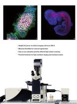
• Amplify the power of confocal imaging with Leica HCS A • Maximum flexibility for universal applications • Easy-to-use automation provides efficient high content screening • Powerful hardware for high resolution imaging and maximum content 2 Leica Design by Christophe
Abrir o catálogo na página 2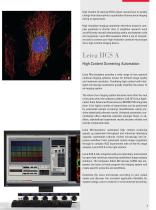
High Content Screening (HCS) allows researchers to quickly change from descriptive to quantitative fluorescence imaging during an experiment. High resolution imaging automation therefore answers complex questions in shorter time. It simplifies research work and efficiently reveals relationships within and between cells and organisms. Leica Microsystems offers a set of innovative tools to convert your high resolution confocal microscope into a high content imaging device. Leica HCS A High Content Screening Automation Leica Microsystems provides a wide range of true spectral confocal imaging...
Abrir o catálogo na página 3
Features • High resolution imaging • Time saving automation • Open architecture • Platform independent results • OME data formats • Perfect integration Modern research is a continuous cycle of experiment design, setup imaging, data handling, and analysis to discover live's processes and answer questions. Power of Imaging Result Content Analysis Data Processing Sample Preparation Automated Leica High Content Screening High resolution imaging techniques answer many questions in modern life science. For high content screening, automation is essential to efficiently achieve results. Intelligent...
Abrir o catálogo na página 4
Sample eparation Leica High Content Screening Automation Data Management Content Analysis Research Result a Rat brain slice, small neuron network layer 5. Interneurons (Alexa 594, red) and Pyramidal Cell Oregon (Bapta 1, calcium sensitive, green). Courtesy of Dr. Thomas Nevian, Institute of Physiology, University of Bern, Switzerland. b Danio rerio – Zebrafish – Nuclear and Acetylated α-Tubulin staining of 6 days flh:eGFP Zebrafish larvae Nuclei (Hoechst, blue), acetylated tubulin (red) and neurons (GFP, green). Courtesy of ICI Imaging Centre IGBMC, Illkirch, France. Choose the adequate...
Abrir o catálogo na página 5
Many companies offer dedicated imaging routines for dedicated assays only. Leica Microsystems provides standard solutions for routine experiments plus maximum flexibility to freely adapt imaging. Features • Smart user interfaces • Workflow oriented wizards • Predefined templates • Easy adjustment • Quick start Easy-to-use AutomationWe Keep It Simple! Wizards guide the user through an experiment in a streamlined way. Design follows function - benefit from clear user interfaces, ensuring fast training and the highest productivity. Predefined Scanning Templates Place the specimen carrier on...
Abrir o catálogo na página 6
Mouse diaphragm muscle stained against neuro-filament 150. Mosaic: xyz: 5 x 5 x 101 images Green: Alexa Fluor 488-secondary antibody and acetylcholine receptors (Red: Alexa Fluor 647-alpha-bungarotoxin). Courtesy of Dr. R. Rudolf, Cellular Signaling in Skeletal Muscle, Karlsruhe Institute of Technology, Germany.
Abrir o catálogo na página 7
Automated High Content Screening Simplifies Da LAS AF software simplifies routines. Leica Microsystems’ goal is to make daily work as easy as possible so researchers can concentrate on the results, not on the imaging process. We Keep it Simple! LAS AF MATRIX Mosaic Fine details as well as an overview are important when evaluating experimental results. Today, simple routine tasks, such as stitching of individual images are challenging. Leica HCS A provides entirely new designed mosaic algorithms for excellent results at the push of a button. Leica LAS AF MATRIX Mosaic generates large high...
Abrir o catálogo na página 8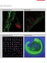
Daily Routines High Resolution Single Image High Content Mosaic Fast Multiwell Plate Screening High Content Information
Abrir o catálogo na página 9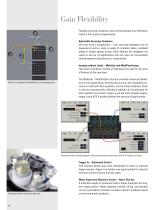
Gain Flexibility Flexible scanning conditions, even on the smallest scan field level, match a full variety of experiments. Adjustable Scanning Templates No need to be a programmer – new scanning templates can be designed at micron scale to easily fit chamber slides, multiwell plates or simple spotter arrays. Once defined, the templates are ready to use for all applications and can even be conveniently shared between laboratories or communities. Imaging without Limits – MultiJob and MultiPositioning Feel free to combine a variety of individual scan jobs for any area of interest of the specimen....
Abrir o catálogo na página 10
Autofocus Routines Five autofocus algorithms are available, optimized for different setups. The suitable routine is selected from a pull-down menu. After the initial scan, the software automatically creates a focus map with true sample topology. This map is used for fast, accurate z-positioning during the scan. According to the size and planarity of the samples, the optimal number and positions of the autofocus points can be freely defined. Z-Drift Compensation Live specimens can grow in long-time measurements, changing the z-position of interest. Microscope conditions can change due to...
Abrir o catálogo na página 11
Automated High Content Screening for Quant Detect Rare Events On-the-Fly! LAS AF MATRIX Developer Suite Highly complex assay designs are now possible with LAS AF MATRIX Developer Suite. The Mitocheck project (1), conducted at the EMBL in Heidelberg, is an excellent example of comprehensive and flexible automation: The self-acting process automates mitosis identification. High Content Screening with Fast Pre-Scan Pre-Scan – Object Identification At first, a fast, low resolution pre-scan of sample plates is routinely performed to identify the events of interest (1). After each scan, OME-image...
Abrir o catálogo na página 12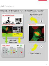
h Interactive System Control: Fully Automated Mitosis Acquisition High Content Scan Feedback Automation Image Analysis embl ijjjjjjjl 1. Pro-Scan 2. Analysis 3. Classification Segmentation Feature Extractor Object Object Selection
Abrir o catálogo na página 13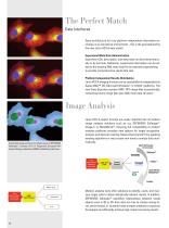
The Perfect MatchData Interfaces Open architecture for truly platform independent information exchange in an interactive environment - this is the goal attained by the new Leica HCS A data model. Experiment Meta Data Administration Experiment IDs, description, and meta data can be entered manually or by barcode. Additional experiment information can be added to the existing XML meta data file by external programming to provide comprehensive result data sets. Platform Independent Results Distribution Leica HCS A imaging formats can be used platform independent on Apple MAC™ OS, Microsoft...
Abrir o catálogo na página 14Todos os catálogos e folhetos técnicos Leica Microsystems
-
 M844 F40/F20
16 Páginas
-
 EnFocus
12 Páginas
-
 M822 F40 / F20
12 Páginas
-
 M320 for ENT
12 Páginas
-
 ARveo 8
16 Páginas
-
 M320 Dental Brochure
12 Páginas
-
 Emspira 3
4 Páginas
-
 Exalta
2 Páginas
-
 FLEXACAM C1
4 Páginas
-
 EM KMR3
8 Páginas
-
 EM UC7
16 Páginas
-
 EM TRIM2
8 Páginas
-
 EM RAPID
8 Páginas
-
 EM ICE
12 Páginas
-
 EM TXP
10 Páginas
-
 EM RES102
12 Páginas
-
 EM TIC 3X
16 Páginas
-
 TL4000 BFDF
16 Páginas
-
 F12 I floor stand
6 Páginas
-
 XL Stand
4 Páginas
-
 TL3000 Ergo & TL5000 Ergo
4 Páginas
-
 KL300 LED
8 Páginas
-
 LED1000
16 Páginas
-
 LED3000 BLI
20 Páginas
-
 LED5000 NVI
20 Páginas
-
 LED3000 NVI
20 Páginas
-
 LED3000 DI
20 Páginas
-
 LED5000 HDI
20 Páginas
-
 LED5000 CXI
20 Páginas
-
 LED2500
8 Páginas
-
 LED5000 MCI
20 Páginas
-
 LED3000 MCI
20 Páginas
-
 LED5000 SLI
20 Páginas
-
 LED3000 SLI
20 Páginas
-
 LED2000
8 Páginas
-
 LED5000 RL
20 Páginas
-
 LED3000 RL
20 Páginas
-
 MZ10 F
4 Páginas
-
 M165 FC
16 Páginas
-
 M205 FCA, M205 FA
16 Páginas
-
 M125 C, M165 C, M205 C, M205 A
12 Páginas
-
 A60 F, A60 S
16 Páginas
-
 M50, M60, M80
12 Páginas
-
 DVM6
DVM616 Páginas
-
 TCS SPE
20 Páginas
-
 DFC450 C
6 Páginas
-
 DFC295
6 Páginas
-
 MC170 HD
6 Páginas
-
 DFC3000 G
6 Páginas
-
 DMC4500
4 Páginas
-
 ICC50 W, ICC50 E
6 Páginas
-
 DFC7000 T, DFC7000 GT
4 Páginas
-
 DFC9000
2 Páginas
-
 IC90 E
6 Páginas
-
 DMC6200
8 Páginas
-
 DMC5400
8 Páginas
-
 SFL7000
4 Páginas
-
 EL6000
4 Páginas
-
 SFL100
4 Páginas
-
 SFL4000
4 Páginas
-
 DMi8 S Platform
2 Páginas
-
 THUNDER Imager Live Cell
2 Páginas
-
 DMi8 M / C / A
12 Páginas
-
 DM IL LED
12 Páginas
-
 DMi1
DMi16 Páginas
-
 DM3 XL
7 Páginas
-
 FS M
FS M4 Páginas
-
 FS C
FS C4 Páginas
-
 FS CB
FS CB4 Páginas
-
 DM3000, DM3000 LED
16 Páginas
-
 DM750 M
12 Páginas
-
 DM750
DM75012 Páginas
-
 DM500
DM50012 Páginas
-
 DM300
DM3008 Páginas
-
 DM12000 M
8 Páginas
-
 DM8000 M
8 Páginas
-
 DM1750 M
12 Páginas
-
 DM4 M, DM6 M
12 Páginas
-
 DM4 P, DM2700 P, DM750 P
12 Páginas
-
 DM2000, DM2000 LED
16 Páginas
-
 DM1000
16 Páginas
-
 DCM8
DCM816 Páginas
-
 DM1000 LED
16 Páginas
-
 DM2500
16 Páginas
-
 DM4 B & DM6 B
16 Páginas
-
 DM6 M LIBS
2 Páginas
-
 S9 Series
12 Páginas
-
 Z6 APO
16 Páginas
-
 Z16 APO
16 Páginas
-
 Leica M530 OHX for ENT
4 Páginas
-
 M620 F20
8 Páginas
-
 M220 F12
8 Páginas
-
 2D and 3D IOL guidance systems
8 Páginas
-
 Leica Application Suite X
4 Páginas
-
 Proveo 8
16 Páginas
-
 M525 F20
12 Páginas
-
 EnVisu Leica Handheld OCT
8 Páginas
-
 PROvido
8 Páginas
-
 Leica M530 OHX
16 Páginas
-
 Leica TCS SP8 STED 3X
24 Páginas
-
 Leica TCS SP8 Objective
24 Páginas
-
 Leica AOBS
16 Páginas
-
 Leica DMC2900
6 Páginas
-
 Leica DMshare
2 Páginas
-
 Leica_DM4000-6000-BrochureTechnical
10 Páginas
-
 Leica_DMshare_ICC50-Flyer_en
2 Páginas
-
 Leica_DMshare_IC80_HD-Flyer_en
2 Páginas
-
 Leica_DMshare_EZ4_HD-Flyer_en
2 Páginas
-
 Leica_DMshare_EC3-Flyer_en
2 Páginas
-
 Leica_SL801-Flyer
2 Páginas
-
 Leica_SCN400-Flyer_Clinical
2 Páginas
-
 DM2700 M
12 Páginas
-
 Leica_SR_GSD_Technical-Brochure
8 Páginas
-
 Leica_SR_GSD-Brochure
10 Páginas
-
 Leica_AF6000-Brochure
16 Páginas
-
 Leica motCorr-Flyer_EN
4 Páginas
-
 Leica TCS SP8-Flyer
2 Páginas
-
 Leica TCS SP8-Brochure
40 Páginas
-
 Leica TCS SP8 X-Flyer
2 Páginas
-
 Leica TCS SP8 Scan Head-Flyer_EN
1 Páginas
-
 Leica TCS SP8 STED-Flyer
2 Páginas
-
 Leica TCS SP8 HyD-Flyer
2 Páginas

