Grupo: Danaher

Excertos do catálogo

Redefine the Limits of Microscopy Widefield super-resolution with ground state depletion
Abrir o catálogo na página 1
Wideeld RCC-FG1 cells, immunouorescence staining against α tubulin with Alexa Fluor® 647. Wideeld Golgi body, B16 (Mouse melanoma cell line), Golgi targeting signal of β 1,4-galactosyltransferase, fused to EYFP. • Resolve more – down to ~ 20 nm • Resolve online – see your super-resolution image build up on screen as it is acquired • Resolve smartly – use standard dyes for highest exibility 2 Courtesy of Prof. Ralf Jacob. Philipps University Marburg, Germany GSD Dr. Yasushi Okada, Department of Cell Biology and Anatomy, Graduate School of Medicine, University of Tokyo, Japan Courtesy of...
Abrir o catálogo na página 2
Courtesy of Prof. Ralf Jacob. Philipps University Marburg, Germany ld GSD Wideeld MDCK cells: Microtubules, Alexa Fluor 642 (red) and TyrMicrotubules, Alexa Fluor® 488 (green). ® Bridging the Two Worlds of Light and Electron Microscopy Fluorescence microscopy has developed into one of the most important tools in life science research. The ultimate aim is to examine single molecule and sub cellular components – structures that are too small to be resolved using standard light microscopy. Pioneers always want to push the limits, striving to learn more and demanding to see further. But when...
Abrir o catálogo na página 3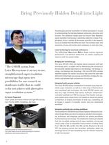
Bring Previously Hidden Detail into Light Visualizing the precise localization of cellular processes is crucial to understanding the interplay between molecules, structures and function. The additional insight given by Ground State Depletion super-resolution microscopy is extremely useful for a range of applications, since a number of structures currently in the research focus are smaller than the diffraction limit. This includes endo- and exosomes, viruses and nuclear pore complexes, to name but a few. Latest technology for maximum performance The SuMo Stage (Suppressed Motion Stage)...
Abrir o catálogo na página 4
New Technology Breaks the Diffraction Barrier How to resolve below the diffraction limit The resolution of a regular fluorescence microscope image is limited by diffraction to approximately half the wavelength of the emitted light. To separate fluorophores that are closer together, the solution is to ensure that not all illuminated fluorophores are able to emit simultaneously. To this end, the excitation light is used such that almost all fluorophores in the samples instantly turn dark. The continuously shining excitation light removes fluorophores from their ground state, leaving only a...
Abrir o catálogo na página 5
Leica SR GSD – The Fast Way to S Your Benets • Maximum resolution down to 20 nm • The SuMo Stage, with Suppressed Motion technology, minimizes drift for accurate localization of molecules • Online super-resolution image projection – see results as they are acquired • Full application exibility offered by combining super-resolution with TIRF and epiuorescence on a multi-purpose live cell imaging system • Standard uorochromes can be used – no need to change protocols • Powerful lasers for the highest exibility in uorochrome selection • Large set of powerful image processing tools 6 Redene the...
Abrir o catálogo na página 6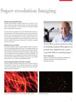
o Super-resolution Imaging Resolve previously hidden details GSDIM super-resolution imaging needs time to acquire a sufcient number of single molecule events. The image acquisition runs in the background – but how long do you collect images before stopping the experiment? To help answer this question, the Leica SR GSD offers online image projection of the super-resolution image. During acquisition, the user sees the image building up online. This feature puts you in full control of the experiment – you can decide to stop or continue the image acquisition to achieve the best result. There is...
Abrir o catálogo na página 7
Technical Specifications Lateral resolution* – Maximum 20 nm – Typical 40 nm Laser – 488 nm/300 mW – 532 nm/500 mW – 642 nm/500 mW – 405 nm/30 mW Imaging modes – GSD super-resolution – TIRF (also available with GSD) – EPI uorescence (also available with GSD) – Brighteld – DIC/PH Laser safety System class 1 Field of view – 18 x 18 μm (GSD mode) – 50 x 50 μm (standard TIRF) Supported dyes – Alexa Fluor® 488 – Rhodamine-6G – Atto 532 and 488 – Alexa Fluor® 532 – Alexa Fluor® 546 – Atto 565 and 568 – Alexa Fluor® 568 – Alexa Fluor® 647 – YFP Imaging Real-time image processing and display of...
Abrir o catálogo na página 8
GSD (Ground State Depletion) Microtubule-staining: anti-p-tubulin/Alexa Fluor® 647 Courtesy: Wernher Fouquet, Leica Microsystems in collaboration with Anna Szymborska and Jan Ellenberg, EMBL, Heidelberg, Germany. Resolving power of different microscopy techniques: The Leica SR GSD is advancing light microscopy to a new level of resolution.
Abrir o catálogo na página 9
The statement by Ernst Leitz in 1907, "With the User, For the User," describes the fruitful collaboration with end users and driving force of nnovation at Leica Microsystems. We have developed five brand values to live up to this tradition: Pioneering, High-end Quality, Team Spirit, Dedication to Science, and Continuous Improvement. For us, living up to these values means: Living up to Life. Leica Microsystems operates globally in three divisions, where we rank with the market leaders. LIFE SCIENCE DIVISION The Leica Microsystems Life Science Division supports the imaging needs of the...
Abrir o catálogo na página 10Todos os catálogos e folhetos técnicos Leica Microsystems
-
 M844 F40/F20
16 Páginas
-
 EnFocus
12 Páginas
-
 M822 F40 / F20
12 Páginas
-
 M320 for ENT
12 Páginas
-
 ARveo 8
16 Páginas
-
 M320 Dental Brochure
12 Páginas
-
 Emspira 3
4 Páginas
-
 Exalta
2 Páginas
-
 FLEXACAM C1
4 Páginas
-
 EM KMR3
8 Páginas
-
 EM UC7
16 Páginas
-
 EM TRIM2
8 Páginas
-
 EM RAPID
8 Páginas
-
 EM ICE
12 Páginas
-
 EM TXP
10 Páginas
-
 EM RES102
12 Páginas
-
 EM TIC 3X
16 Páginas
-
 TL4000 BFDF
16 Páginas
-
 F12 I floor stand
6 Páginas
-
 XL Stand
4 Páginas
-
 TL3000 Ergo & TL5000 Ergo
4 Páginas
-
 KL300 LED
8 Páginas
-
 LED1000
16 Páginas
-
 LED3000 BLI
20 Páginas
-
 LED5000 NVI
20 Páginas
-
 LED3000 NVI
20 Páginas
-
 LED3000 DI
20 Páginas
-
 LED5000 HDI
20 Páginas
-
 LED5000 CXI
20 Páginas
-
 LED2500
8 Páginas
-
 LED5000 MCI
20 Páginas
-
 LED3000 MCI
20 Páginas
-
 LED5000 SLI
20 Páginas
-
 LED3000 SLI
20 Páginas
-
 LED2000
8 Páginas
-
 LED5000 RL
20 Páginas
-
 LED3000 RL
20 Páginas
-
 MZ10 F
4 Páginas
-
 M165 FC
16 Páginas
-
 M205 FCA, M205 FA
16 Páginas
-
 M125 C, M165 C, M205 C, M205 A
12 Páginas
-
 A60 F, A60 S
16 Páginas
-
 M50, M60, M80
12 Páginas
-
 DVM6
DVM616 Páginas
-
 HCS A
HCS A20 Páginas
-
 TCS SPE
20 Páginas
-
 DFC450 C
6 Páginas
-
 DFC295
6 Páginas
-
 MC170 HD
6 Páginas
-
 DFC3000 G
6 Páginas
-
 DMC4500
4 Páginas
-
 ICC50 W, ICC50 E
6 Páginas
-
 DFC7000 T, DFC7000 GT
4 Páginas
-
 DFC9000
2 Páginas
-
 IC90 E
6 Páginas
-
 DMC6200
8 Páginas
-
 DMC5400
8 Páginas
-
 SFL7000
4 Páginas
-
 EL6000
4 Páginas
-
 SFL100
4 Páginas
-
 SFL4000
4 Páginas
-
 DMi8 S Platform
2 Páginas
-
 THUNDER Imager Live Cell
2 Páginas
-
 DMi8 M / C / A
12 Páginas
-
 DM IL LED
12 Páginas
-
 DMi1
DMi16 Páginas
-
 DM3 XL
7 Páginas
-
 FS M
FS M4 Páginas
-
 FS C
FS C4 Páginas
-
 FS CB
FS CB4 Páginas
-
 DM3000, DM3000 LED
16 Páginas
-
 DM750 M
12 Páginas
-
 DM750
DM75012 Páginas
-
 DM500
DM50012 Páginas
-
 DM300
DM3008 Páginas
-
 DM12000 M
8 Páginas
-
 DM8000 M
8 Páginas
-
 DM1750 M
12 Páginas
-
 DM4 M, DM6 M
12 Páginas
-
 DM4 P, DM2700 P, DM750 P
12 Páginas
-
 DM2000, DM2000 LED
16 Páginas
-
 DM1000
16 Páginas
-
 DCM8
DCM816 Páginas
-
 DM1000 LED
16 Páginas
-
 DM2500
16 Páginas
-
 DM4 B & DM6 B
16 Páginas
-
 DM6 M LIBS
2 Páginas
-
 S9 Series
12 Páginas
-
 Z6 APO
16 Páginas
-
 Z16 APO
16 Páginas
-
 Leica M530 OHX for ENT
4 Páginas
-
 M620 F20
8 Páginas
-
 M220 F12
8 Páginas
-
 2D and 3D IOL guidance systems
8 Páginas
-
 Leica Application Suite X
4 Páginas
-
 Proveo 8
16 Páginas
-
 M525 F20
12 Páginas
-
 EnVisu Leica Handheld OCT
8 Páginas
-
 PROvido
8 Páginas
-
 Leica M530 OHX
16 Páginas
-
 Leica TCS SP8 STED 3X
24 Páginas
-
 Leica TCS SP8 Objective
24 Páginas
-
 Leica AOBS
16 Páginas
-
 Leica DMC2900
6 Páginas
-
 Leica DMshare
2 Páginas
-
 Leica_DM4000-6000-BrochureTechnical
10 Páginas
-
 Leica_DMshare_ICC50-Flyer_en
2 Páginas
-
 Leica_DMshare_IC80_HD-Flyer_en
2 Páginas
-
 Leica_DMshare_EZ4_HD-Flyer_en
2 Páginas
-
 Leica_DMshare_EC3-Flyer_en
2 Páginas
-
 Leica_SL801-Flyer
2 Páginas
-
 Leica_SCN400-Flyer_Clinical
2 Páginas
-
 DM2700 M
12 Páginas
-
 Leica_SR_GSD_Technical-Brochure
8 Páginas
-
 Leica_AF6000-Brochure
16 Páginas
-
 Leica motCorr-Flyer_EN
4 Páginas
-
 Leica TCS SP8-Flyer
2 Páginas
-
 Leica TCS SP8-Brochure
40 Páginas
-
 Leica TCS SP8 X-Flyer
2 Páginas
-
 Leica TCS SP8 Scan Head-Flyer_EN
1 Páginas
-
 Leica TCS SP8 STED-Flyer
2 Páginas
-
 Leica TCS SP8 HyD-Flyer
2 Páginas

