 Website:
Leica Microsystems
Grupo: Danaher
Excertos do catálogo

Leica Objectives Superior Optics for Confocal and Multiphoton Research Microscopy
Abrir o catálogo na página 1
Leica Microsystems: Optics for Your Discoveries We have designed and produced superior optics for a wide variety of applications in research, industry, and medicine for more than 160 years. Today, the innovation power of our optics esigners and the experience and d expertise of our precision optical engineers come together to provide microscopes with the best possible optics for spectral imaging. A sophisticated state-of-the-art production process yields objectives that deliver superla Working “with the user, for the user” (Ernst Leitz I, 1843–1920) tive image quality. We also help you...
Abrir o catálogo na página 2
AOBS – First reprogrammable beam splitter Leica TCS SP5 X – First white light laser confocal with tunable VIS excitation Leica TCS SP5 – Broadband confocal with the first switchable tandem scanner λ blue objectives optimized for 405 nm excitation Leica TCS SP8 – New confocal platform, modular system Leica TCS SP5 MP with OPO, excitation up to 1300 nm First motCORR objectives with motorized correction collar IRAPO objectives with color correction up to 1300 nm Leica TCS SP8 STED 3X – Fast and direct super-resolution HC PL APO STED WHITE with superior broadband color correction Leica HC PL...
Abrir o catálogo na página 3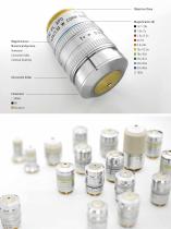
Numerical Aperture Correction Collar Confocal Scanning Correction Collar Objective Class
Abrir o catálogo na página 4
Choose the Best Objective for Your Needs The name of an objective describes its specifications and target applications. This guide gives you an overview of the technical terms and abbreviations. PL/PLAN – Excellent field planarity (1) Objective with a flattened field of view for the representation of thin specimens, crucial for confocal microscopy of thin objects. APO/IRAPO/FLUOTAR – Appropriate color correction is required for colocalization in multicolor specimens. Wavelength range of correction (2) In addition, high transmission of an objective for the excitation and emission wavelengths...
Abrir o catálogo na página 5
High Numerical Aperture for Best Resolution Numerical aperture and wavelength directly influence the resolving power of a microscope. Resolution improves with higher numerical apertures and lower wavelengths. NUMERICAL APERTURE POINT SPREAD FUNCTION The numerical aperture (NA) of an objective is described by The point spread function (PSF) describes how an imaging the sine of the half-angle α of the maximum cone of light that system represents a point object in three dimensions. The PSF can enter or exit the lens multiplied by the refractive index n of a fluorescence microscope is dependent...
Abrir o catálogo na página 6
Rayleigh Limit LATERAL RESOLUTION AXIAL RESOLUTION AND OPTICAL SECTION THICKNESS For a rough estimation of the resolving power of a fluores- The volume of the PSF is not only restricted horizontally in cence microscope in x and y, applying the Rayleigh criterion the focus plane but also vertically along the optical axis of is usually sufficient. Here, the maximum of the Airy disk of the microscope (z). The axial resolution of a microscope system one point overlaps with the first minimum of the Airy disk is worse than its lateral resolution, approximately by a factor of the second point...
Abrir o catálogo na página 7
Highly Corrected Optics for Better Images Precise optical design and high manufacturing standards ensure that the imaging errors inherent in every optical system are reduced to a minimum. SPHERICAL ABERRATIONS FIELD CURVATURE Spherical aberration is the dominant Field curvature is a monochromatic imaging error that needs to be corrected aberration that causes the optimal focus in high numerical aperture optics. position to vary with the image point An objective with spherical aberrations position. It increases quadratically with the distance between the image point and the center of field....
Abrir o catálogo na página 8
AXIAL COLOR LATERAL COLOR Axial color causes the optimal focus Lateral chromatic aberration causes the position to vary with the wavelength. magnification to vary with the This aberration occurs in the center of wavelength. It increases linearly with field and remains constant over the the distance between the image point whole field of view. In polychromatic applications this Image points near the edge of the field aberration causes a loss of contrast, show a distinctive colored smearing for colored fringes and a best focus which the human eye is very sensitive. In quantitative...
Abrir o catálogo na página 9
It’s All About Color Preparing your specimen for microscopy is only the first step to a stunning image. Choosing the best objective for your purpose should be done with as much care. Besides magnification and numerical aperture, the appropriate color correction is crucial. Apochromatic Color Correction Apochromats allow the image resolution to reach the diffraction limit over a wide range of wavelengths. For fluorescence imaging, 1.0 all excitation and detection wavelengths must be included to ensure that image resolution is as high as physically possible. For optimal colocalization,...
Abrir o catálogo na página 10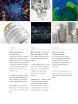
IR Color Correction for Multicolor Confocal Scanning Multiphoton Imaging and CARS Fluorescence Imaging The apochromatic Leica CS2 objectives Leica PL IRAPO objectives are highly are optimized for confocal scanning specified for improved multiphoton PL FLUOTAR objectives feature good (CS). Their color correction is outstanding imaging. chromatic correction for visible over the whole field of view for precise wavelengths. This makes them well colocalization of different fluorophores. The IR apochromats are color corrected suited for standard fluorescence In particular the lateral color has...
Abrir o catálogo na página 11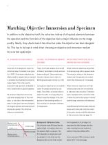
Matching Objective Immersion and Specimen In addition to the objective itself, the refractive indices of all optical elements between the specimen and the front lens of the objective have a major influence on the image quality. Ideally, they should match the refractive index the objective has been designed for. This has to be kept in mind when choosing an objective and immersion medium for a certain application. OIL: STANDARD FOR FIXED SAMPLES GLYCEROL: THE OPTIMUM FOR MOUNTED WATER: PERFECT MATCH FOR LIVE CELL IMAGING AND THICK SPECIMENS Immersion oil is designed to match the Today, most...
Abrir o catálogo na página 12Todos os catálogos e folhetos técnicos Leica Microsystems
-
M844 F40/F20
16 Páginas
-
EnFocus
12 Páginas
-
M822 F40 / F20
12 Páginas
-
M320 for ENT
12 Páginas
-
ARveo 8
16 Páginas
-
M320 Dental Brochure
12 Páginas
-
Emspira 3
4 Páginas
-
Exalta
2 Páginas
-
FLEXACAM C1
4 Páginas
-
EM KMR3
8 Páginas
-
EM UC7
16 Páginas
-
EM TRIM2
8 Páginas
-
EM RAPID
8 Páginas
-
EM ICE
12 Páginas
-
EM TXP
10 Páginas
-
EM RES102
12 Páginas
-
EM TIC 3X
16 Páginas
-
TL4000 BFDF
16 Páginas
-
F12 I floor stand
6 Páginas
-
XL Stand
4 Páginas
-
TL3000 Ergo & TL5000 Ergo
4 Páginas
-
KL300 LED
8 Páginas
-
LED1000
16 Páginas
-
LED3000 BLI
20 Páginas
-
LED5000 NVI
20 Páginas
-
LED3000 NVI
20 Páginas
-
LED3000 DI
20 Páginas
-
LED5000 HDI
20 Páginas
-
LED5000 CXI
20 Páginas
-
LED2500
8 Páginas
-
LED5000 MCI
20 Páginas
-
LED3000 MCI
20 Páginas
-
LED5000 SLI
20 Páginas
-
LED3000 SLI
20 Páginas
-
LED2000
8 Páginas
-
LED5000 RL
20 Páginas
-
LED3000 RL
20 Páginas
-
MZ10 F
4 Páginas
-
M165 FC
16 Páginas
-
M205 FCA, M205 FA
16 Páginas
-
M125 C, M165 C, M205 C, M205 A
12 Páginas
-
A60 F, A60 S
16 Páginas
-
M50, M60, M80
12 Páginas
-
DVM6
16 Páginas
-
HCS A
20 Páginas
-
TCS SPE
20 Páginas
-
DFC450 C
6 Páginas
-
DFC295
6 Páginas
-
MC170 HD
6 Páginas
-
DFC3000 G
6 Páginas
-
DMC4500
4 Páginas
-
ICC50 W, ICC50 E
6 Páginas
-
DFC7000 T, DFC7000 GT
4 Páginas
-
DFC9000
2 Páginas
-
IC90 E
6 Páginas
-
DMC6200
8 Páginas
-
DMC5400
8 Páginas
-
SFL7000
4 Páginas
-
EL6000
4 Páginas
-
SFL100
4 Páginas
-
SFL4000
4 Páginas
-
DMi8 S Platform
2 Páginas
-
THUNDER Imager Live Cell
2 Páginas
-
DMi8 M / C / A
12 Páginas
-
DM IL LED
12 Páginas
-
DMi1
6 Páginas
-
DM3 XL
7 Páginas
-
FS M
4 Páginas
-
FS C
4 Páginas
-
FS CB
4 Páginas
-
DM3000, DM3000 LED
16 Páginas
-
DM750 M
12 Páginas
-
DM750
12 Páginas
-
DM500
12 Páginas
-
DM300
8 Páginas
-
DM12000 M
8 Páginas
-
DM8000 M
8 Páginas
-
DM1750 M
12 Páginas
-
DM4 M, DM6 M
12 Páginas
-
DM4 P, DM2700 P, DM750 P
12 Páginas
-
DM2000, DM2000 LED
16 Páginas
-
DM1000
16 Páginas
-
DCM8
16 Páginas
-
DM1000 LED
16 Páginas
-
DM2500
16 Páginas
-
DM4 B & DM6 B
16 Páginas
-
DM6 M LIBS
2 Páginas
-
S9 Series
12 Páginas
-
Z6 APO
16 Páginas
-
Z16 APO
16 Páginas
-
Leica M530 OHX for ENT
4 Páginas
-
M620 F20
8 Páginas
-
M220 F12
8 Páginas
-
2D and 3D IOL guidance systems
8 Páginas
-
Leica Application Suite X
4 Páginas
-
Proveo 8
16 Páginas
-
M525 F20
12 Páginas
-
EnVisu Leica Handheld OCT
8 Páginas
-
PROvido
8 Páginas
-
Leica M530 OHX
16 Páginas
-
Leica TCS SP8 STED 3X
24 Páginas
-
Leica AOBS
16 Páginas
-
Leica DMC2900
6 Páginas
-
Leica DMshare
2 Páginas
-
Leica_DM4000-6000-BrochureTechnical
10 Páginas
-
Leica_DMshare_ICC50-Flyer_en
2 Páginas
-
Leica_DMshare_IC80_HD-Flyer_en
2 Páginas
-
Leica_DMshare_EZ4_HD-Flyer_en
2 Páginas
-
Leica_DMshare_EC3-Flyer_en
2 Páginas
-
Leica_SL801-Flyer
2 Páginas
-
Leica_SCN400-Flyer_Clinical
2 Páginas
-
DM2700 M
12 Páginas
-
Leica_SR_GSD_Technical-Brochure
8 Páginas
-
Leica_SR_GSD-Brochure
10 Páginas
-
Leica_AF6000-Brochure
16 Páginas
-
Leica motCorr-Flyer_EN
4 Páginas
-
Leica TCS SP8-Flyer
2 Páginas
-
Leica TCS SP8-Brochure
40 Páginas
-
Leica TCS SP8 X-Flyer
2 Páginas
-
Leica TCS SP8 Scan Head-Flyer_EN
1 Páginas
-
Leica TCS SP8 STED-Flyer
2 Páginas
-
Leica TCS SP8 HyD-Flyer
2 Páginas


