
Excertos do catálogo
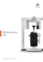
Thinking ahead. Focused on life.
Abrir o catálogo na página 1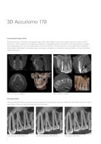
Unsurpassed image clarity The 3D Accuitomo 170 offers unsurpassed image clarity. With 9 fields of view and multiple acquisition modes, the 3D Accuitomo 170 can meet all of your diagnostic needs with unparalleled quality. Its super-fine minimal voxel size of just 80µm allows diagnosing even the most subtle details of bone and dentition. The 3D Accuitomo 170 is highly recommended by leading dental radiologists for periodontology, oral surgery, endodontics, orthodontics, dental implants, for the maxillofacial region and beyond. Stunning clarity The high resolution 80μm acquisitions provide...
Abrir o catálogo na página 2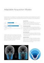
Adaptable Acquisition Modes The 3D Accuitomo 170 is equipped with four imaging modes that allow flexibility when scanning patients with a variety of diagnostic needs and clinical indications. Choose High Resolution and High Fidelity modes for the best quality images, or High Speed for patients that have difficulty remaining still. The 3D Accuitomo 170 will adapt to suite your diagnostic and your patients’ needs. Standard Mode 360º scan 17.5 sec, 180º scan: 9 sec Standard mode offers images of exceptional clarity and is suitable for limited and wide views of temporal bone, paranasal, sinus,...
Abrir o catálogo na página 3
Small Fields of View Stay focused on your region of interest by selecting the best field of view for your indication. Volume diameters as small as 40 mm or as large as 100 mm can be selected for the dentition.
Abrir o catálogo na página 4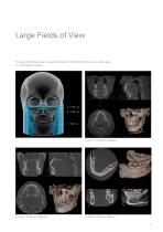
Large Fields of View For larger maxillofacial scans, select a diameter of ø100 to ø170 to cover a wider range of maxillofacial surgeries.
Abrir o catálogo na página 5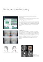
Simple, Accurate Positioning The three positioning laser beams and an LCD make patient positioning easy. The chinrest stabilizes the patient`s head to avoid movement. Scout images enable even more accurate positioning. Easy as One, Two, Three. First, the patient`s initial position is set and recorded using the three positioning laser beams. Then, the region of interest is aligned in the LCD. The chair automatically moves into the optimal position. During the X-ray exposure, the patient is stabilized by the chinrest and the headrest. 2–direction scout For even more accurate positioning,...
Abrir o catálogo na página 6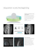
Acquisition is only the beginning 180 degree reconstruction to reduce motion artifacts Did the patient move during acquisition? Do you have to scan them again? Not any more! The 3D Accuitomo 170 allows you to extract 1/2 of a 360° scan at any point to remove that initial jolt or that unintended swallow at the end of the acquisition. (For 360 ° acquisitions only) Remove unintended motion artifacts 180° Patient moved 360° Scan Extract 180° motion-free data 360° scan exhibiting patient movement artifacts 180° of motion-free data extracted. Zoom reconstruction The Accuitomo series is equipped...
Abrir o catálogo na página 7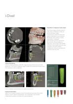
Acquisition to diagnosis made simple The i-Dixel imaging software offers a wide variety of features to help you quickly and easily create comprehensive treatment plans and explain those plans to your patients. Mandibular canal marking, implant presentation, multiplanar reconstruction are just a few of the features that i-Dixel provides for diagnoses. i-Dixel is also fully DICOM compliant and provides quick and easy integration with both practice management software and advanced treatment planning tools. Volume Rendering Volume rendering displays a solid 3D image showing the bone structure...
Abrir o catálogo na página 8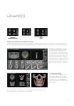
Freedom from platform and simplicity of design i-Dixel WEB runs as a web service on an X-ray server PC included with your Morita X-ray system. It serves as a local and secure web-based dental image processing service that you can access throughout your practice on a wide range of devices. No software installation needed With the latest advancements in web technology, i-Dixel WEB gives you the freedom to view your images wherever you want and however you want. Gone are the days of complicated chairside PC setups, and limited choices of hardware. Mac OS X and even iPads can be used to view...
Abrir o catálogo na página 9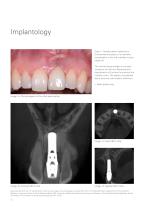
Implantology Case 1: Female patient referred for 3-dimensional analysis of an esthetic complication in the left maxillary incisor region (a). The clinical status exhibits a mucosal recession as well as a flattening and discoloration of the facial mucosa at the implant crown. The patient complained about recurrent peri-implant infections. * : Nasal palate tube Image 1a: Clinical aspect at the initial examination Image 1c: Axial CBCT slice Image 1b: Coronal CBCT slice Image 1d: Sagittal CBCT slice Case courtesy Prof. em. Dr. Daniel Buser, Clinic for oral surgery and somatology University Bern...
Abrir o catálogo na página 10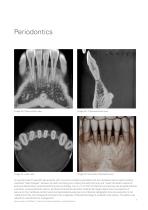
Image 2a: Para-coronal view Image 2b: Cross-sectional view Image 2c: Axial view Image 2d: Volumetric Rendered view An asymptomatic 51-year-old male presents with numerous complaints associated with the mandibular anterior teeth including unesthetic “black triangles” between the teeth, shrinking gums making the teeth look long, and “loose”The patient reports an . extensive dental history of periodontal bone loss and therapy. A 4 cm x 4 cm FOV at 0.08 nominal voxel size was acquired and paracoronal (a), cross-sectional (b), axial (c), and three dimensional volumetric rendered (d) images...
Abrir o catálogo na página 11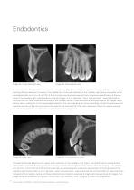
Image 3a: Cross-sectional view Image 3b: Para-sagittal view An asymptomatic 27-year-old female presents immediately after fixed orthodontic appliance therapy with bitewing imaging showing diffuse radiolucent increase in the middle third of the pulp chamber of the maxillary right canine compared to the contralateral side. An 8 cm x 8 cm FOV at 0.25 nominal voxel size was acquired and a suspicious opacification of the pulp canal of the maxillary right canine noted on axial (a) images. A 4 cm diameter “zoom reconstruction “was performed at a nominal 0.08 mm voxel resolution centered on the...
Abrir o catálogo na página 12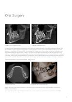
Oral Surgery Image 5a: MPR Panoramic view Image 5b: Para-sagittal view An asymptomatic 56 year-year-old male presents with a history of incidental discovery of possible mandibular pathology in the left mandible on routine panoramic imaging taken 1 month previously. A 10 cm x 10 cm FOV at 0.25 nominal voxel size was acquired, A reformatted MPR panoramic (a), and para-sagittal (b) images clearly show a single, well-defined, corticated, irregularly-shaped bilobular low density lesion anterior to the lingula and mandibular foramen within the left ascending ramus with extension inferiorly...
Abrir o catálogo na página 13Todos os catálogos e folhetos técnicos Morita
-
 Tri Auto ZX2+
8 Páginas
-
 Instrument brochure
16 Páginas
-
 EndoWave Feilen
2 Páginas
-
 Signo T100
12 Páginas
-
 Signo Z300
12 Páginas
-
 Signo T500
13 Páginas
-
 Signo G10 II
41 Páginas
-
 Root ZX mini
2 Páginas
-
 Veraviewepocs 2D
10 Páginas
-
 Veraview IC5 HD
5 Páginas
-
 GUMMETAL®
5 Páginas
-
 Lubrina 2
8 Páginas
-
 Veraviewepocs 3D R100
16 Páginas
-
 AdvErL EVO
6 Páginas
-
 TriAuto mini
10 Páginas
-
 Implant Dentistry
36 Páginas
-
 i-Dixel - 2D & 3D Imaging Software
2 Páginas
-
 Veraview X800
10 Páginas
-
 TorqTech handpieces
2 Páginas
-
 DentaPort ZX with OTR function
4 Páginas
-
 Veraview iX
2 Páginas
-
 EndoWave OTR Sequence FM
2 Páginas
-
 Genuine Root ZX
2 Páginas
-
 i-Dixel Web
2 Páginas
Catálogos arquivados
-
 Soaric for endodontics
2 Páginas
-
 Soaric
19 Páginas
-
 PenCure 2000
2 Páginas
-
 Proven Vacuum Mixing Technology
2 Páginas
-
 3D Accuitomo Clinical Case Evidence
40 Páginas
-
 Brochure "Endodontics is an Art"
10 Páginas