
Excertos do catálogo
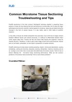
Common Microtome Tissue Sectioning Troubleshooting and Tips Paraffin sectioning is the most common histological technique applied in preparing tissue sections. It does not only observe the form and structure of normal cells, but it is also the major pathological and forensic scientific method applied for studying, observing and determining the changes in the form of cellular tissues. It is also widely used in other fields of scientific research. Living cells or tissues are mostly transparent and colourless. Due to the lack of image contrast between different tissues and cellular structures, it is difficult to differentiate between them under regular glass lens. Tissues die fast and decay after leaving the organism, losing its original structure. Therefore, in order to get a clear image of their forms and structures, tissues have to be fixated, embedded, cut and dyed in preparation of sample sections. Paraffin sectioning involves steps including sampling, fixation, rinsing and dehydration, waxing, embedding, cutting and mounting, dewaxing, staining, dehydration, clearing, mounting and so on. Cutting is one of the most crucial parts that determines the section results. There’re lots of factors affecting the microtome tissue sectioning performance. What are the common problems of microtome tissue sectioning? How to solve these problems? Crumbled Ribbon
Abrir o catálogo na página 1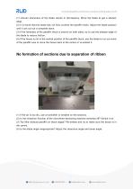
(1) Uneven sharpness of the blade results in discrepancy. Move the blade to get a sharper edge. (2) It is found that the blade has not fully covered the paraffin block. Adjust the blade position until it can cut out a complete block. (3) If the hardness of the paraffin block is uneven on both sides, try to use the sharper edge of the blade to reduce friction. (4) If the tissue is not in the central position of the paraffin block, use the blade to cut out some of the paraffin wax to move the tissue back to the centre or re-embed it. No formation of sections due to separation of ribbon (1) If...
Abrir o catálogo na página 2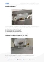
Rolled-up Section (1) Is the paraffin block too cold? Increase the temperature accordingly. Use a brush to spread out the section and press it firmly while cutting it into 2 to 3 flattened slices. (2) Paraffin block is too hard? Add softer wax. (3) The blade is too blunt? Change the blade. (4) The bevel angle is too big? Narrow it down. Ribbons crumble and stick on the knife (1) Is the temperature of the block too high? Lower the temperature appropriately. s the paraffin block too soft? Freeze it more. -3-
Abrir o catálogo na página 3
(2) A thin layer of paraffin remains on top of the blade? Clean the blade and scrub the excessive paraffin wax on top. Use Xylene to clean the blade. (3) Blunt blade? Get a new blade. (4) Too many fats? Remove the fats during preparation and squeeze out the fats at the stage of embedding. (1) The knife is chipped? Move the blade. (2) Impurities in paraffin wax? Remove the impurities and get high-quality paraffin. (3) Excess wax remains on the blade? Clean the blade. (4) The tissue is too hard? Soak it in the water to soften it. (5) Hemostats inside the tissue? Take them out. Train lines on...
Abrir o catálogo na página 4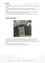
(1) Are the clamps and other parts of the machine getting loose? Make sure everything is firmly locked. (2) The blade bevel angle is too wide? Narrow it down to an appropriate degree. (3) The specimen extends too much? Retract it appropriately and move the knife holder forward. (4) The paraffin block is not firmly locked? Clean the paraffin block and the cassette clamp. (5) The tissue is too hard? Decalcify its surface and cut it slowly. (6) Are there worn microtome parts? Maintenance or machine replacement is necessary. Uneven Thickness (1) Are there worn microtome parts? Maintenance or...
Abrir o catálogo na página 5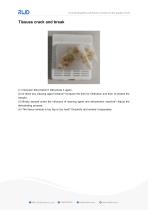
Tissues crack and break (1) Improper dehydration? Dehydrate it again. (2) Is there any clearing agent residue? Increase the time for infiltration and then re-embed the sample. (3) Brisky sample under the influence of clearing agent and dehydration machine? Adjust the dehydrating process. (4) The tissue sample is too big or too hard? Decalcify and embed it separately.
Abrir o catálogo na página 6Todos os catálogos e folhetos técnicos RWD Life Science
-
 R821 Fiber photometry Brochure
6 Páginas
-
 RWD Catalog for Cell& Molecular Biology
12 Páginas
-
 RWD Catalogue for Life Science Research
46 Páginas
-
 RWD Gradient Thermal Cycler
2 Páginas
-
 RWD M1324 High-Speed Microcentrifuge
2 Páginas
-
 Rotary Microtomes S710
2 Páginas
-
 RM500/RM600 Veterinary Monitor
2 Páginas
-
 RWD Nanoliter micro-injector R480
2 Páginas
-
 RWD Osmotic infusion Pump
2 Páginas
-
 RWD Syringe Pump R462
1 Páginas
-
 RWD Rotating impactor 68099Ⅱ
2 Páginas
-
 RWD Microcentrifuge M1324R
2 Páginas
-
 RWD Veterinary anesthesia R640
2 Páginas
-
 RWD Infusion imager RFLSI Ⅲ
2 Páginas
-
 RWD Mice stereotaxic frame 68001
1 Páginas
-
 RWD Versatile Animal ICU R-CU510
2 Páginas
-
 RWD Surgical Instruments
18 Páginas
-
 RWD Microtome Cryostat FS800A FS800
2 Páginas
-
 RWD Veterinary medical equipment
32 Páginas
-
 RWD C100 Automated cell counter
2 Páginas
-
 RWD R-CU510 Versatile Animal ICU
2 Páginas
-
 RWD D-pro Veterinary Dental Unit
2 Páginas
-
 RWD R415 Animal Ventilatorr
1 Páginas
-
 MP-500 Micropipette Electrode puller
2 Páginas


Catálogos arquivados
-
 PRODUCT CATALOGUE 2017
51 Páginas
-
 2015 Product Catalogue
44 Páginas
