
Excertos do catálogo

Swept Source Optical Coherence Tomography
Abrir o catálogo na página 1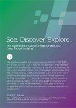
See. Discover. Explore. The diagnostic power of Swept Source OCT Deep Range Imaging¹. “Swept Source adds a new dimension to OCT. The TOPCON DRI Swept Source OCT is easy to use, provides unique clinical information, and has improved my practice. For the first time, we can in-vivo visualize not only the vitreo-retinal interface but also the cortical vitreous which is important at the time when more and more therapies are delivered via intra-vitreal injections. Deeper imaging brings choroidal thickness, helping guide my clinical decisions. Seeing more helps guide my therapy and allows me to...
Abrir o catálogo na página 2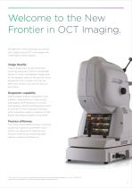
Welcome to the New Frontier in OCT Imaging. The DRI OCT Triton combines the world’s first2 Swept Source OCT technology with multimodal fundus imaging. Image Quality Triton’s Swept Source with its 100 kHz scanning speed and 1,050nm wavelength results in a clear and detailed images even for the deepest layers of the eye with short acquisition time. Visualize not only the retina and vitreous, but also the choroid and sclera1. Diagnostic capability Seeing deeper makes it possible to have a better understanding of many ocular pathologies1.With features such as OCT angiography, fundus...
Abrir o catálogo na página 3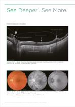
Proliferative diabetic retinopathy Lateral: 12mm Courtesy: Prof. P. E. Stanga, Manchester Royal Eye Hospital, Manchester Vision Regeneration (MVR) Lab at N IHR/ Welcome Trust Manchester CRF & University of Manchester Courtesy: Prof. P. E. Stanga, Manchester Royal Eye Hospital, Manchester Vision Regeneration (MVR) Lab at N IHR/ Welcome Trust Manchester CRF & University of Manchester * FA photography and FAF photography can be performed using only DRI OCT Triton Plus. 1) Fabio Lavinsky, Daniel Lavinsky. Novel perspectives on swept‑source optical coherence tomography. Int J Retin Vitr (2016)...
Abrir o catálogo na página 4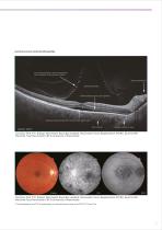
Central serous chorioretinopathy Lateral: 12mm Courtesy: Prof. P. E. Stanga, Manchester Royal Eye Hospital, Manchester Vision Regeneration (MVR) Lab at N IHR/ Welcome Trust Manchester CRF & University of Manchester Courtesy: Prof. P. E. Stanga, Manchester Royal Eye Hospital, Manchester Vision Regeneration (MVR) Lab at N IHR/ Welcome Trust Manchester CRF & University of Manchester * FA photography and FAF photography can be performed using only DRI OCT Triton Plus.
Abrir o catálogo na página 5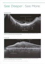
Pathological myopia Lateral: 12mm Courtesy: Prof. P. E. Stanga, Manchester Royal Eye Hospital, Manchester Vision Regeneration (MVR) Lab at N IHR/ Welcome Trust Manchester CRF & University of Manchester Macular pucker Lateral: 12mm Courtesy: Prof. P. E. Stanga, Manchester Royal Eye Hospital, Manchester Vision Regeneration (MVR) Lab at N IHR/ Welcome Trust Manchester CRF & University of Manchester 1) Fabio Lavinsky, Daniel Lavinsky. Novel perspectives on swept‑source optical coherence tomography. Int J Retin Vitr (2016) 2:25
Abrir o catálogo na página 6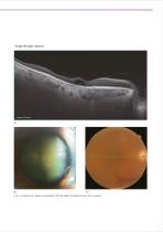
Image through cataract a, b, c courtesy of Kazuya Yamagishi, MD (Hirakata Yamagishi Eye Clinic, Japan)
Abrir o catálogo na página 7
Discover What Lies Beneath TOPCON’s SS OCT AngioTM combines OCT angiography with a Swept Source OCT. OCTARATM, a proprietary image processing algorithm, provides highly sensitive angiographic detection3, allowing for visualization of vascular structures even in the choroid and deeper retinal layers. High-sensitivity Imaging and Deeper Intravascular Flow Visualization1 Swept Source technology and OCTARATM allow the deeper structures to be visualized with less depthdependent signal roll-off 3. Additionally, the 1μm wavelength makes OCT Angiography imaging possible for patients with media...
Abrir o catálogo na página 8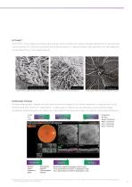
OCTARATM OCTARATM is the image processing technology which extracts the signal changes derived from vascular flow using multiple OCT B-Scans acquired at the same position. It demonstrates high sensitivity for the detection of low blood flow in microvasculature3. Choroid (GA ) (3.0 x 3.0 mm) Courtesy of SriniVas R. Sadda, M.D., Courtesy: SriniVas R. Sadda, M.D., Doheny Eye Institute, UCLA Doheny Eye Institute, UCLA Choroid (CNV ) Courtesy: SriniVas R. Sadda, M.D., Doheny Eye Institute, UCLA RPC(Glaucoma) Courtesy: Kazuya Yamagishi, MD., Hirakata Yamagishi Eye Clinic, Japan Multimodal Viewing...
Abrir o catálogo na página 9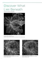
Discover What Lies Beneath Proliferative diabetic retinopathy SS OCT AngioTM Montage Courtesy: Akihiro Ishibazawa, MD, PhD. Asahikawa Medical University Graduate School of Medical Sciences, Hokkaido, Japan Before treatment Courtesy: Akihiro Ishibazawa, MD, PhD. Asahikawa Medical University Graduate School of Medical Sciences, Hokkaido, Japan After treatment Courtesy: Akihiro Ishibazawa, MD, PhD. Asahikawa Medical University Graduate School of Medical Sciences, Hokkaido, Japan
Abrir o catálogo na página 10
Branch retinal vein occlusion Courtesy:Yuichiro Ogura, MD, Professor and Chairman, Department of Ophthalmology and Visual Science, Nagoya City University, Nagoya, Japan
Abrir o catálogo na página 11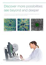
Discover more possibilities: see beyond and deeper OCTA metrics on Triton SS OCT Angio allows clinicians to objectively and quantitatively assess retinal vasculature, providing valuable insights into the patient’s eye health. Courtesy: Michael H. Chen, O.D.
Abrir o catálogo na página 12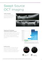
Swept Source OCT Imaging 1,050nm wavelength The longer wavelength light provides better tissue penetration, allowing visualization into the deepest layers of the eye1. Courtesy: Professor Jose Maria Ruiz Moreno MD, University of Albacete, Spain Swept Source OCT technology; scanning speed of 100,000 A-scans/sec The fast scanning speed of 100,000 A-scans/sec enables capture of clear B-scans4 by acquiring more A-scans within a given image acquisition involuntary eye movements such as saccades time. This helps to reduce artifacts from Invisible scan lines The invisible 1,050nm wavelength light...
Abrir o catálogo na página 13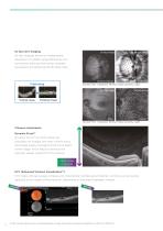
Projection image en face imaging allows for independent dissection of a depth range defined by two boundaries, selected from seven possible boundaries, by flattening the 3D data cube. Lamina Cribosa Original image Lamina Cribosa Courtesy: Prof. T. Nakazawa, MD,PhD, Tohoku University, Japan Flattened image Courtesy: Prof. T. Nakazawa, MD,PhD, Tohoku University, Japan Vitreous visualization Dynamic FocusTM Dynamic FocusTM on Triton allows for acquisition of images with near uniform focus and image quality throughout the entire depth of the image, which helps to enhance the typically weaker...
Abrir o catálogo na página 14Todos os catálogos e folhetos técnicos Topcon Healthcare
-
 HARMONY
7 Páginas
-
 OMS-800 Series
8 Páginas
-
 Aladdin
11 Páginas
-
 CV-5000PRO
10 Páginas
-
 SOLOS
SOLOS4 Páginas
-
 CL-300
6 Páginas
-
 TRK-2P
8 Páginas
-
 KR-1W
KR-1W7 Páginas
-
 KR-1
KR-18 Páginas
-
 KR-800S
10 Páginas
-
 KR-800PA
2 Páginas
-
 KR-800A
6 Páginas
-
 KR-800/RM-800
4 Páginas
-
 Chronos
8 Páginas
-
 ACP-8
ACP-84 Páginas
-
 CC series
5 Páginas
-
 ATE Series
10 Páginas
-
 FS-1 series
4 Páginas
-
 IS-1 Series
16 Páginas
-
 IS-600 III
12 Páginas
-
 IS-100
8 Páginas
-
 SP-1P
SP-1P8 Páginas
-
 Henson 9000
9 Páginas
-
 CT-1P
CT-1P10 Páginas
-
 CT-800A
6 Páginas
-
 CT-800
4 Páginas
-
 MYAH
MYAH8 Páginas
-
 CA-800
9 Páginas
-
 Topcon Slit Lamp Series
7 Páginas
-
 TRC-50DX
5 Páginas
-
 Signal
2 Páginas
-
 TRC-NW8 series
8 Páginas
-
 TRC-NW400
8 Páginas
-
 Maestro2
16 Páginas
Catálogos arquivados
-
 VT-10 Vision Tester
4 Páginas
-
 VT-10
VT-104 Páginas